A 73 year old woman with over a decade old MAGUS develops lymphoma without a Show!
She came to her dentist for residual tooth extraction. Doctors diagnosed her with IgM kappa monoclonal gammopathy (MAGUS) 13 years ago. She had been on a regular follow-up for her disease ever since. At the time of her dental appointment, her antibody levels came out 13.16 g/L. She did not have any autoimmune disease manifestation or history. The good thing was, her disease followed a very silent course. During the entirety of her follow-up period, she never displayed any abnormalities on her X-rays, CT scans, MRIs and even on her PET scan. However, this time it was destined to be a little different.
Digital Palpation Reveals a Lesion
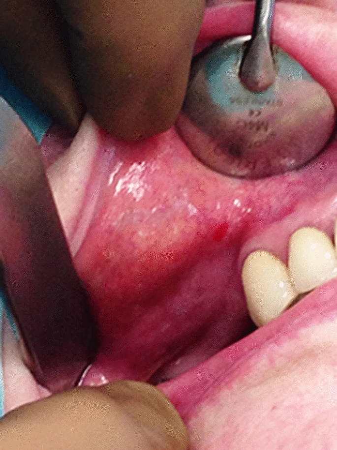
Before extracting her residual root, doctors performed routine inspection and examination. Intraoral inspection did not reveal anything significant. The patient did not have any mucosal tears or ulcers nor did she have any swelling or pain. On palpation however, doctors felt a sub-mucosal nodule on the patient’s right cheek just below the Stenon’s canal opening. The nodule felt firm in consistency. However, it did not elicit any pain.
Doctors decided to surgically excise the nodule along with her residual tooth extraction procedure. They successfully excised the nodule under local anesthesia. And then sent it for histopathological examination. Results dropped some interesting findings. The yellowish-brown nodule consisted of lymphocytic proliferations. It had follicles with germinal centers with intervening lymphomatous infiltrate. These tumor cells displayed plasma cell differentiation in some areas, too. On immunohistochemistry, the cells expressed CD20, CD79a, CD5, Bcl2 and a multitude of other receptors and markers. Thus referring to these findings, doctors labelled the excised nodule as mucosa associated lymphoid tissue (MALT) lymphoma.
MAGUS and MALT: What Are These?
Monoclonal gammopathy of undetermined significance (MAGUS) describes a condition where the body produces abnormal amounts of immunoglobulins (M proteins) from a single clone of plasma cells (monoclonal). These are released into the serum and manifest as an “M spike” on electrophoresis. It differs from another disease that shows an M spike on serum electrophoresis called multiple myeloma where the spike represents an elevated IgG or IgA rather than IgM. Anyhow, MAGUS seldom presents with notable symptoms. It does not cause any organ damage and most of the times, it is discovered only incidentally.
MAGUS, although asymptomatic most of the times, can develop complications such as coagulopathies and autoimmune disturbances, essentially due to the monoclonal antibodies. It can affect kidneys to cause monoclonal gammopathy of renal significance, in which case the manifestations involve renal function disturbances. Furthermore, MAGUS can sometimes progress to develop diseases such as multiple myeloma and other lymphoproliferative disorders. However, the risk remains very low especially for something like a MALT lymphoma.
MALT Lymphoma of the Oral Cavity: What Are the Odds?
MALT lymphoma constitutes one of the subtypes of Non-Hodgkin’s lymphoma that develops extranodally and involves B cells of the marginal zone. It usually develops in people above 50 to 60 years of age and is related to a focus of infection or an autoimmune trigger. The pathogenesis involves proliferation of lymphoid tissue associated with the mucosa of different parts of the body. The commonest site of involvement come out to be gut, more precisely ileum, that has abundant lymphoid tissue in the form of Peyer’s patches. Also, it develops frequently in the stomach where the most common cause comes out to be an Helicobacter pylori infection.
The clinical features of a MALT lymphoma depend upon the site of its occurrence for example dyspepsia in case of gastric involvement. However, some common presentations irrespective of the site subsume fever, weight loss and anemia etc. Anyhow in our patient, doctors discovered the lymphoma in the oral cavity which by all standards is very rare. To top it off, it did not cause any symptoms due to which it remained hidden for a long time. Doctors only caught it while doing a detailed palpation of the oral cavity prior to the patient’s residual tooth extraction procedure.
Management of Our Patient with MAGUS and MALT Lymphoma
After successfully excising the lymphoma, doctors referred the patient to hematology department. There the patient underwent further evaluation for any treatment indication. However since the patient was asymptomatic, doctors only continued her on follow-up without any specific treatment. Fortunately, her IgM kappa levels remained harmless and her lymphoma did not recur.


