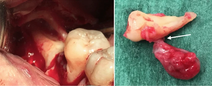
This article describes the case of a patient who developed a lateral periodontal cyst in relation to her impacted right lower third molar tooth.
The 54-year-old woman presented with a complaint of pain in the lower right side of her face. She was healthy and did not have a very significant history otherwise. Her oral examination revealed a swelling in her right lower molar region along with a horizontally impacted third molar. Doctors did periodontal probing on which they found a gingival sulcus 9 millimetres in depth, with purulent material oozing out from it. Hence, they made an initial diagnosis of a gingival abscess.
Radiography Reveals Lateral Periodontal Cyst
On an orthopantomogram, the swelling displayed a radiolucent nature. Its association with the third molar suggested a dentigerous cyst. Hence, the doctors ruled out the gingival abscess and planned their treatment based on their new diagnosis.
As a treatment plan, doctors irrigated the gingiva with chlorhexidine and started the patient on antibiotics and analgesics. They also discharged the patient with advice to rinse the area with chlorhexidine twice a day. After a week, they called the patient and removed the cyst along with the impacted molar and sent the specimen for histopathology. Results confirmed a lateral periodontal cyst. Doctors kept the patient on a follow-up to monitor her for any recurrence. Fortunately, everything went well and the disease did not develop again.
Lateral Periodontal Cyst (LPC): A sneak peek
LPC is a developmental cyst that originates in the teeth. It develops between two teeth or on the lateral side of an erupted tooth. Most commonly, it involves the molars. Doctors often confuse it with a gingival cyst because of a similar point of origin and histopathological characteristics. However, gingival cyst develops in the gums outside the teeth.
LPC shares a long list of differential diagnoses because potentially, all of them manifest as a radiolucent lesion on an orthopantomogram. Thus, narrowing down the list is quite a hustle. First on the list comes a dentigerous cyst (DC). It develops in the region of the third molar or maxillary canine and is taken up on a radiograph as a well-circumscribed radiolucent lesion. However, unlike LPC which can develop as a multifocal lesion, it only develops univocally. Moreover, it comes with features of sclerosis around the crown which we normally do not see in LPC.
The second important differential is a radicular cyst. It closely resembles a lateral periodontal cyst because most of the time, it too develops on the lateral aspect of the tooth. However, it affects an infected non-vital tooth and thus presents with features of inflammation which are never seen with LPC.
The third important differential that doctors should rule out is a glandular odontogenic cyst. It also develops in the teeth in relation to its lateral aspect. But it usually grows aggressively and has crypts within the epithelial cells on histology. Moreover, it also contains ciliated cells which we do not see in LPC.
LPCs: How Do They Originate?
Different researchers give different theories to explain the origin of lateral periodontal cysts. According to some, the cyst develops from remnants of the dental lamina which are found in abundance near the facial faces of the teeth. This explains why these cysts grow laterally. Others suggest that these cysts develop as a result of desquamation from the teeth’ enamel. The peeled-off portions remain latent for some time and then develop into a cyst. This school of thought gets its support from similarities between histological findings of enamel epithelium and that of the cyst.
Another popular theory that explains the origin says that the cyst develops from epithelial cells in the periodontal ligament. Anyhow, the cyst usually follows a benign course and can be removed following dental surgery. The chances of recurrence are low too.
A Rare Presentation
This case represents a rare presentation of LPC in association with impacted teeth. Normally, it develops in association with erupted teeth. Nevertheless, it is important to pick it up on radiology and plan accordingly. Those with impacted teeth can be treated with surgical removal along with the removal of impacted teeth as in our patient. The results are almost always encouraging.


