- The incidence of craniomaxillofacial injuries in the general population because of interpersonal violence has increased significantly over the years.
- Women victims contribute to 20 to 25% of the cases of interpersonal violence and is a leading cause of trauma in females.
- In this case the patient suffered a sharp assault by a sickle by her husband in a drunken state.
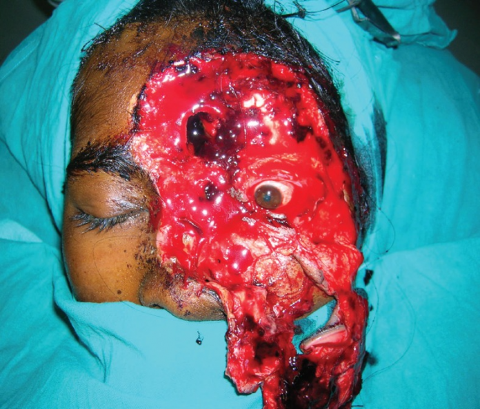
A 25-year-old female patient presented to the emergency with hemifacial avulsion and degloving. The patient had allegedly been assaulted by the husband with a sickle over the left side of the face. Examination showed avulsions and degloving of the left side of the face including the nose, skin on the forehead, left side of the eyebrow, lower eyelids and part of the cheek. Extraocular movements were present with the globe and eyesight intact.
The wound was evaluated under anaesthesia which showed a loss of anterior cortex of the frontal sinus and nasal bones. The part of the frontal sinus was attached to the degloved skin flap, whereas, on the left side, a part of the frontal bone was exposed. Contamination of the wound with small amount of mud particles was also present.
The wound was thoroughly debrided and the devitalised tissue was excised. The degloved flap was attempted to be repositioned to their respective positions. The avulsed structures were relocated with intricate suturing to their positions. The eyelids were relocated with medial and lateral cathopexies under appropriate tension.
Hemifacial Avulsion Degloving Recovery
In addition, there was a significant loss of tissue in the left forehead, over the nasal dorsum and area of the cheek. The mucosa of the exposed frontal sinus was curretted and the cavity was plugged with the outer cortex of the frontal sinus. Additionally, a forehead flap was raised and transposed medially to cover the exposed frontal sinus and frontal bone based on the right supraorbital and supratrochlear vessels. A nasolabial flap was used to cover the defect of skin on the dorsum and right lateral wall of the nose. A split skin graft was grafted at a small defect on the cheek region.
After four weeks of the procedure, a costal cartilage graft was used for augmentation of the columella and dorsum. This was done via a small stab incision over the columella. Moreover, thinning of the nasolabial flap was also done. The postoperative course of the patient’s was quite uneventful.

References
Face Avulsion and Degloving https://www.ncbi.nlm.nih.gov/pmc/articles/PMC4236979/


