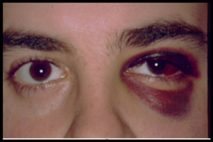
A 21-year-old male presented to the emergency department with a sudden loss of vision in his left eye after a craniofacial trauma. He told the doctors that he had a bicycle accident and wasn’t wearing a helmet. The doctors performed a general physical examination which revealed the subconjunctival haemorrhage along with periorbital ecchymosis in his left eye. Moreover, the patient had an afferent pupillary defect which causes pupillary dilation. However, he was alert and oriented otherwise.
On further evaluation, the visual acuity test showed normal vision in the right eye but the vision of the left eye was severely impaired. The doctors advised for fundoscopy which revealed a well-perfused retina and normal optic disc.
Comminuted Fractures of the Left Optic Canal
The doctors performed radiographic imaging which showed comminuted fractures of the left optic canal in addition to fractures of the floor, medial and lateral walls of the left orbit. It also revealed the fracture of maxillary and sphenoid sinuses. Moreover, the two displaced bone fragments of the sphenoid bone were impinging directly upon the optic nerve.
Management
Doctors started him on intravenous methylprednisolone 1g for 3 consecutive days but it had no improvement on the patient’s condition. Therefore, the patient underwent optic nerve decompression surgery through the endoscopic endonasal approach. They removed the bone fragments which were impinging on the optic nerve. As a result, the total visual acuity of the patient’s left eye returned to normal without any complications
Traumatic Optic Neuropathy
This case highlights the successful management of traumatic optic neuropathy. Although surgery and steroids are usually successful in treating patients with traumatic optic neuropathy, their use is controversial. There is no management protocol for optic neuropathy that exists. Therefore, the management is based on the nature and extent of the injury.
Reference: Cureus


