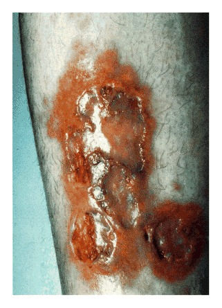
The clinical image shows a lesion on the anterior surface of the lower leg of a 20-year-old patient. The woman’s medical history was consistent with poorly controlled diabetes mellitus. Examination showed atrophic, shiny, yellow-brown telangiectatic plaques, measuring 9 by 5 cm with ulceration and scarring. Doctors diagnosed the patient with necrobiosis lipoidica. She was treated with whirlpool therapy, occlusive dressing, aspirin, pentoxifyline and topical steroids.
What causes necrobiosis lipodica?
Although the cause of the condition is unknown, there are theories that it may be associated with changes in the blood vessel, for example, diabetes and antibody-mediated vasculitis. It begins as a dull, red papules or plaques on the shin which enlarge into one or more yellowish- brown patches with a red rim. The patches are often seen on both shins are rarely found on other sites. On examination the lesions are tender, and have a round, oval and an irregular shape. Other signs include central atrophy, shiny, pale and thinned appearance with prominent blood vessels. In some cases, the patient may also complain of reduced sweating and sensation.
Females are three times more likely to be diagnosed with necorbiosis lipoidica compared to males. It usually develops in young- and middle-aged adults. Statistics show that 1% of patients with diabetes will develop necrobiosis lipoidica. Similarly, it can occur in both diabetes type 1 and type 2. 11% to 65% of patients diagnosed with necrobiosis lipoidica are either diabetic or pre-diabetic. Patients with type 1 diabetes are likely to be younger than patients without diabetes or with diabetes type 2. Other conditions the lesion is associated with include thyroid disease, dyslipidaemia, hypertension and obesity. It is rarely found in children.
Complications of necrobiosis lipoidica
1/3rd of the cases are complicated because of ulcerations, usually followed by a minor injury to an established patch. The ulcer is either very painful or painless. Ulcers because of the associated with the condition are at a risk of secondary bacterial infection and delayed healing. Patients with squamous cell carcinoma may also develop necrobiosis lipoidica.
Source: NEJM


