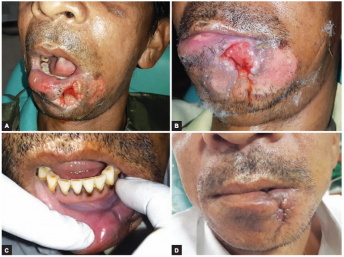
A 43-year-old male presented to the outpatient clinic with a through and through ulcer on the lower lip. The ulcer had appeared a week ago, whereas, the patient gave a history of binge drinking 10 days earlier. The excessive drinking made him dizzy and he fell face first, injuring his lower lip. Moreover, he got the wound sutured at a local hospital.
However, 3 days later, gaping and swelling developed on the site of the wound. Similarly, According to his relatives, worms could be seen falling from the wound. The patient had a poor built and appeared to be mentally unsound. Local examination of the wound revealed a necrotic ulcer (4×3) with undermined edges involving the full thickness of the lower lip. In addition, multiple crevices infested with larvae could be seen within the wound.
On examination, the surrounding mucosa appeared inflamed and tender on palpation associated with bleeding and pus. The pus was sent for culture and sensitivity. Other laboratory investigations including liver function tests and blood investigations were under normal limit with negative serostatus.
Differential diagnoses
Differential diagnoses tungiasis, foreign body reaction, cellulitis and squamous cell carcinoma were considered. However, the presence of larvae was consistent with the diagnosis of oral myiasis.
The maggots were manually removed from an area deep within the wound and a sterile gauze impregnated with turpentine oil was placed at the orifice. The lesion was biopsied to rule out the possibility of carcinoma. There were no evident carcinomatous features or dysplastic changes. Patient was prescribed cefixime and metronidiazole for 3 days as prophylaxis. However, anti-parasitic drugs were not included in the treatment regime. It took 2 consecutive days with exploration, curettage, betadine and warm saline irrigation for the larvae to be completely removed.
The maggots were whitish, without any obvious body processes and 12 to 15 mm long, identified as Chrysomya bezziana species.
The patient was advised chlorohexidine mouthwash for 10 days. Swelling reduced significantly after a week and healthy granulation tissue was seen. The wound had healed completely at 1-month follow-up.
References
Oral Myiasis—A Pauper’s Affection: Case Reports and a Review of 62 Cases https://www.researchgate.net/publication/316905642_Oral_Myiasis-A_Pauper’s_Affection_Case_Reports_and_a_Review_of_62_Cases


