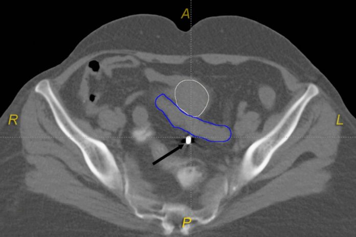
This article highlights the case of uterine perforation during a seemingly straightforward procedure in a cervical-carcinoma patient. The procedure involved placing necessary applicators through the uterine cavity into the left ovoids into the lateral vaginal fornices.
The patient was diagnosed with cervical carcinoma and was referred for intracavitary brachytherapy (ICBT)
He was taken for a CT scan for dose-optimised three dimensional conformal brachytherapy with central tandem. While the patient was undergoing the scan it was seen that the central tandem had pierced through the posterior wall of the uterus entering the abdominal cavity. Other findings included the uterine body as anteflexed. However, the previous examination did not visualise this. The doctors removed the applicators and postponed the procedure.
For successful radiotherapy of cervical carcinoma, it is often essential to integrate brachytherapy with external beam radiotherapy. Brachytherapy facilitates adequate dosing of the cervix, upper-vagina and uterus. Whereas, at the same time it spares the urinary bladder and rectum from excessive dosing. According to the case study, “ICBT is a time-tested treatment for cervical carcinoma, wherein the accessible utero-cervical cavity is irradiated from-within by the placement of applicators to facilitate radioactive-source loading”.
Even though ultrasound-guidance is known for reducing the risk of uterine perforations, a majority of clinicians have not adopted this method for applicator placement
There are several benefits of using applicators which the study has also highlighted. For example, in the current era, the applicators are loaded with computer-controlled iridium-192 source which delivers the radiation. After the tandem and ovoids are placed, the dose stimulation can be done with a traditional x-ray stimulation or with CT-stimulation. Although using x-ray stimulation is less expensive and also simpler, CT-stimulation is clinically advantageous. This enables volumetric dose-optimisation. As with this case, it can also help with detecting an unexpected uterine perforation.
Source: BMJ


