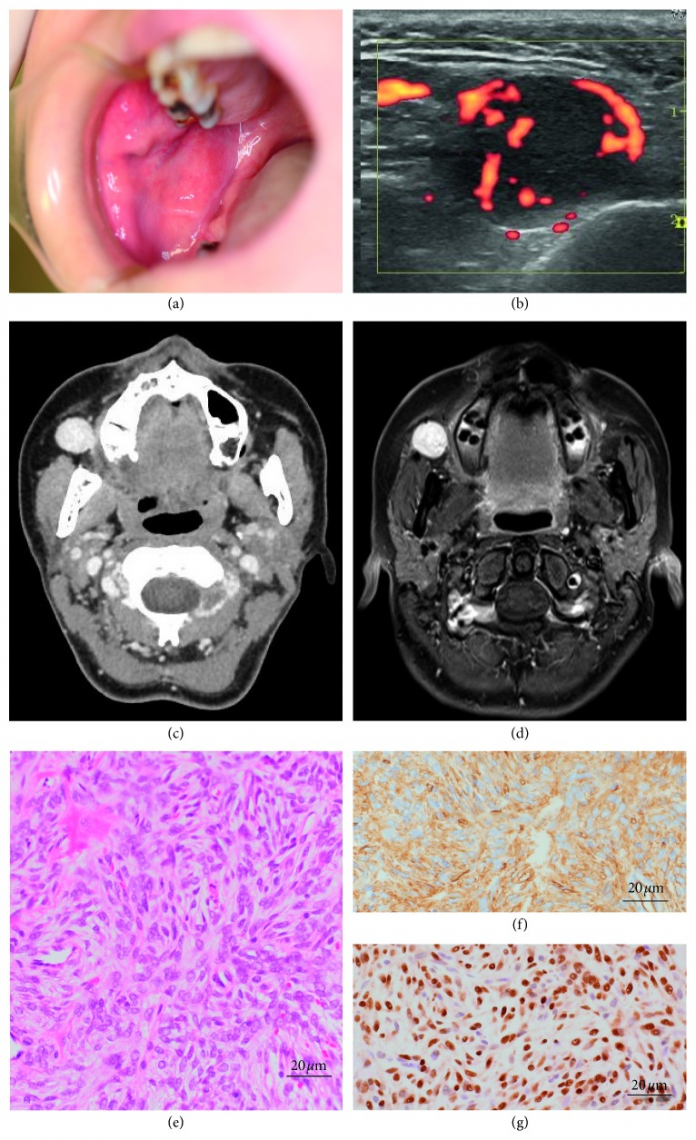
Case of solitary fibrous tumour in buccal space in 39-year-old.
This article describes the case of a solitary fibrous tumour in the buccal space of a 39-year-old Japanese woman. The patient presented to the Oral and Maxillofacial Surgery department at Okoyama University Hospital with a 3-year history of a buccal mass. The mass had been growing slowly for 3 years. However, there were no medical records regarding the mass. Clinical examination showed a well-defined mass with rounded margins in the buccal space, measuring 1.5 x 1.5 cm. The mass was free from skin and muscles.
Examination did not show any palpable lymph nodes in the neck. Neither did the mass cause any pain. Doctors advised a colour doppler ecographic examination which showed a high flow velocity of the blood surrounding the mass. Contrast-enhanced CT, T1 weighted image on magnetic resonance imaging revealed a 1.5 x 1.5 cm homogenous mass, present in front of the masseter muscle. The mass showed well-defined margins. Fine-needle aspiration biopsy showed spindle cells arranged and lined in a patternless manner. The spindle cells were strongly positive for CD34. STAT 6 and vimentin on immunohistochemistry. However, negative for EMA, S100, and α-SMA.
Doctors removed the mass surgically under general anaesthesia.
The mass was surgically removed under general anaesthesia. The tumour was encapsulated with connective tissue and was easily separated from the later structure. Doctors discharged the patient 4 days after surgery. There were no signs of nerve injury or recurrence at 12 months follow-up.
References
Solitary Fibrous Tumor Arising in the Buccal Space https://www.ncbi.nlm.nih.gov/pmc/articles/PMC6803731/


