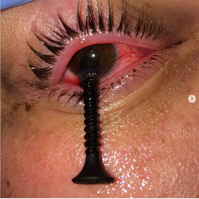
This patient while mounting frames fell victim to a misdirected screw that got embedded in the cornea, therefore the patient was brought to the emergency room for medical attention to remove the screw. Physical examination of the eye revealed a large screw in the infratemporal region of the cornea accompanied by surrounding oedema.
Ophthalmic emergency rooms reflect figures of around 38-52% ocular trauma and out of them, 0.9-1.8% need inpatient hospital care.
The incidence of ocular trauma is particularly high in young males aged between 18 to 25 years, owing to work, assaults, accidents, sports, etc. Although the majority of these cases are preventable provided that suitable steps are taken to avoid such debilitating injuries. With an emphasis on the ergonomics, certain professions are provided with instructions of protective eyewear but availability remains a concern. Moreover, in children of the developing countries, the most common cause of preventable, unilateral blindness is ocular trauma.
Certain tests are essentially required for a thorough examination of the eye.
Checking visual acuity, using the Snellen chart, instantly after eye injury is indispensable. In such cases, information regarding pre-injury visual acuity helps in determining the extent of the damage.
Also, patients should be evaluated for lens dislocation using a direct ophthalmoscope and slit-lamp examination is done to look for penetration of the anterior chamber and corneal perforation. Seidel test aids in detecting aqueous humour leakage; negative tests are suggestive of partial-thickness injury
Absolutely no pressure should be applied on the globe in hope of self-healing.
CT scan is the investigation of choice for precisely localizing orbital and intraocular foreign body and skull X-ray can be done to detect radio-opaque foreign bodies and also to exclude associated fractures of cranium and face. In cases of the ruptured globe, ultrasound can be used.
A point noteworthy here is that magnetic resonance imaging (MRI) is absolutely contraindicated for the metallic intraocular foreign body as the movement of the embedded metal due to the magnetic field of MRI can cause further intraocular damage. In cases where no foreign body is observed in the eye but the patient persistently complains of foreign body sensation and discomfort then a foreign body is presumed until proven otherwise.
Surgical management is directed towards repair of corneal laceration and foreign bodies’ removal. The aim of management is to restore normal anatomy by creating a watertight wound and by minimizing scarring.
In the initial 3 weeks, rapid recovery is expected whereas between 4 to 12 weeks, a final prediction of visual acuity. In the following period of up to 3 years, delayed changes can be seen.
The patient in the discussion here underwent a post-operative Seidel test which was negative with a slightly displaced pupil. His recent visual acuity was 20/50 and he had a visible cataract. After removal of K sutures, 1-2 months were given to the cornea to heal prior to cataract surgery.
References:
John R Acerra, M. (2019, September 03). Globe Rupture. Retrieved from Medscape: https://emedicine.medscape.com/article/798223-overview
Aghadoost D. (2014). Ocular trauma: an overview. Archives of trauma research, 3(2), e21639. https://doi.org/10.5812/atr.21639


