
A unit for burn and reconstructive surgery received a 30-year-old patient with an abnormal scalp appearance since his puberty. Physical examination revealed convoluted folds and creases spanning the entire vertex and the upper occipital region. Temporal regions were spared. All routine laboratory tests were within the normal range, suggesting a diagnosis of primary essential cutis vertices gyrata (CVG). The patient was presented with a surgical option of expanding the affected part.
Cutis verticis gyrata (CVG), also called paquidermia verticis gyrata, cutis verticis plicata, and bulldog scalp syndrome, is a term used to describe a scalp condition with crests and trough appearance of the scalp resembling a cerebriform pattern with thickened scalp skin. CVG has been classified into primary and secondary forms, with the former having no known cause while the latter can be due to diseases like acanthosis nigricans, diabetes, cretinism, connective tissue disorders, graves disease, dermatological inflammatory conditions like eczema, psoriasis, dermatitis, etc., amyloidosis, leukaemia, Turner syndrome, posttraumatic, etc.
Primary CVG affects postpubertal males hinting towards the role of testosterone, whereas secondary CVG may be present at birth or according to the disease that is associated with it. 0.5% of patients with CVG have found to be mentally retarded.
Cutis verticis gyrata has a propensity towards the vertex and the occipital region, seldom it may affect the entire scalp. The affected regions, when examined, reveal anterior to posterior, soft, boggy and spongy folds that remain fixed i.e. unchanged even after pressure or traction. The overlying skin of these 2-12 folds remains unchanged but the density of hair is sparse over the folds and normal in the furrows. If the furrows get secondarily affected by infections, maceration and malodorous odour may be present.
If CVG is present at birth or is associated with other abnormalities, MRI or CT scan may be warranted and if the affected newborn is a female, then the chromosomal analysis is done to exclude Turner syndrome. EEG is done to evaluate the accompanying mental disorder if present.
Primary CVG may be treated symptomatically or surgically. Counseling regarding local hygiene is essential. Surgery is often performed for psychological or cosmetic reasons.
Surgical options include:
- Total resection followed by grafting
- Tissue expander under healthy skin followed by delayed grafting
- Partial resection of the lesion.
The resected sample is sent for histopathology which is usually normal in primary CVG cases. Some samples may show thickened collagen, hypertrophied pilosebaceous glands and multiple sweat glands. The histopathological results of secondary CVG are governed by the underlying disease.
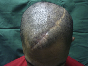
The patient in discussion here underwent surgery for insertion of a 500-ml rectangular tissue expander in the vertex. The postoperative period remained uneventful. Over the next 2 months, expander inflation was performed in the outpatient department twice every week. After 2 months, the folds over the scalp had flattened so the expander was removed and excess deformations were excised.
At the 18th month follow-up, there was a significant improvement.
References:
Yang, J. J., Sano, D. T., Martins, S. R., Tebcherani, A. J., & Sanchez, A. P. (2014). Primary essential cutis verticis gyrata – case report. Anais brasileiros de dermatologia, 89(2), 326–328. https://doi.org/10.1590/abd1806-4841.20142949
Garden JM, Robinson JK. Essential primary cutis verticis gyrata. Treatment with the scalp reduction procedure. Arch Dermatol. 1984 Nov. 120(11):1480-3. [Medline].
Mishra A, Tehrani H, Hancock K, Duncan C. Management of primary cutis verticis gyrata with tissue expansion and hairline lowering foreheadplasty. J Plast Reconstr Aesthet Surg. 2010 Feb 1. [Medline].
Zhao D, Li J, Wang K, Guo X, Lang Y, Peng L, et al. Treating cutis verticis gyrata using skin expansion method. Cell Biochem Biophys. 2012 Mar. 62(2):373-6. [Medline].


