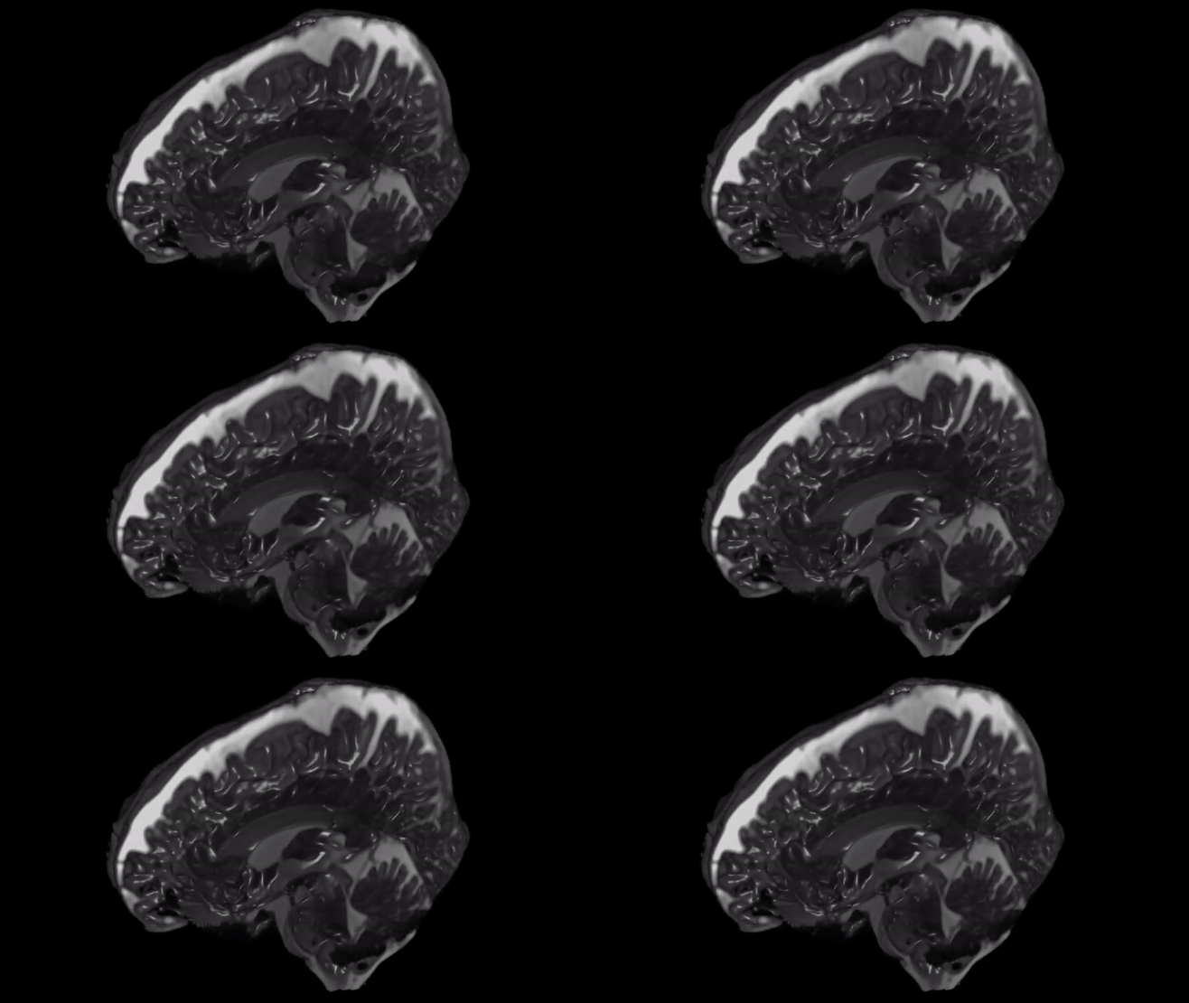
3D aMRI produces 3D footage of the human brain as fluid flows around it
A video showing how the brain wobbles each time blood and other fluids pass through the organ recently surfaced the internet. The incredible phenomenon was captured using a brain scanning technique called 3D aMRI. The 3D image of the human brain shows how fluid flows through and around it.
Two recent studies used a brain-scanning technique to capture real time 3D videos of the brain. The video shows brain tissues pulsating in response to blood rushing through the blood vessels and cerebrospinal fluid. The purpose of the video is to amplify and exaggerate movements in the brain for easy analysis. The motion is very small and at most between 0.002 inches and 0.015 inches. However, the imaging amplifies it 25 times, making it easier for researchers to assess the motion with better detail, direction and amplitude.
The new scanning technique paves way for more accurate diagnosis and treatment approaches of diseases in which fluid is blocked from flowing through the brain.
One such condition is hydrocephalus, in which there is an accumulation of cerebrospinal fluid within the brain. This causes an increase in pressure inside the skull, according to Samantha Holdsworth, co-author on both studies. “We’ve got a lot of work to do to really prove its clinical application … but that’s the nature of all new technology,” she said. “We’re just sort of at the beginnings of what can be achieved.”
The new scanning technique was created with basic MRI using strong magnets for applying a magnetic field in the body. As a response, the hydrogen nuclei lined up within this field. However, when the current is switched off, depending on the tissue that surround then, the nuclei snap back into position at different rates. There are several other MRI techniques that can also be used to track motion in the brain, such as, Displacement Encoding with Stimulated Echoes (DENSE) and phase-contrast MRI. “The advantage of the amplified MRI is that you can see the motion in relation to the underlying anatomy, which is this really exquisite anatomy”, said Holdsworth.
Researchers are currently using this technique to study Chiari I malformation, in which the section of the brain containing the cerebellum is small or deformed. Therefore, it puts pressure on and crowds the brain. Holdsworth is also using this technology to scan brains of patient with concussions to evaluate how fluid flows through their brains after injury.
References
See how the brain wobbles with each heartbeat in incredible new videos https://www.livescience.com/3D-footage-of-brain-in-motion.html?fbclid=IwAR2iQ_ghPE76FYDYUu2nQtrpiedZj5z4vxj3OveXtKWxaefhLYLMTjKiN34


