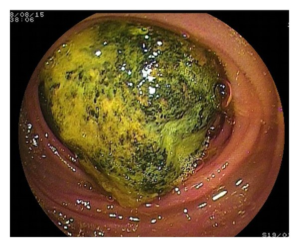
Case of 69-year-old with small bowel obstruction
Small bowel obstruction is usually caused because of a concretion of foreign material found in the stomach, and it is rarely caused by bezoars. It is commonly seen in patients who have undergone gastric surgery. Bezoars are a solid mass of indigestible material which accumulated in the digestive tract. The bezoars usually form in the stomach, sometimes in the small intestine and rarely in the large intestine. They can occur in both children and adults.
The bezoars usually get impacted in the narrowest portion of the small bowel. This article describes the case of a 69-year-old Japanese man who was admitted to the hospital with epigastraligia (pain in the epigastric region). The patient was diagnosed with small bowel bezoars after a double balloon enteroscopy (DBE) was performed. DBE allows for complete visualisation of the small bowel bezoars and is also used for therapeutic interventions.
The patient’s physical examination was consistent with the findings of generalised distension and diffuse tenderness.
There were no visible signs of peritoneal distension. His medical history revealed that he had recently eaten a persimmon, which if eaten frequently can lead to the formation of a persimmon bezoar. A bezoar is an indigestible mass that consists of fibres, skins and seeds trapped in the gastrointestinal system.
Doctors advised laboratory tests which showed an elevated white cell count and C-reactive protein. All other tests including liver function test and chemistry were normal. An erect x-ray of the abdomen was consistent with small bowel gas. A contrast examination of the ileus tube was also performed which showed that ileum was totally obstructed because of the mass. For further evaluation, a computed tomography was performed. The findings showed a dilated small bowel loop with a mass measuring 4 cm. 7 days after the patient’s presentation, a retrogade DBE was performed which a hard mass blocking the distal ileum. The bezoars, although, could not be fragmented.
Doctors operated on the bezoar impacting the ileum via an extraction enterotomy. It was located 100 cm proximal to the ileocecal valve. Doctors discharged the patient in a satisfactory condition, 9 days after the operation.
Source: NCBI


