
According to the case report published in The Journal of Emergency Medicine, a 17-year-old boy accidentally swallowed a small sewing pin which ended up piercing his heart.
The teenager had presented to the emergency department with a three-day history of chest pain. It was described as a sharp, substernal chest pain that radiated to the back and got aggravated on lying down or deep inspiration. He had also reported having episodes of nausea, vomiting, and diarrhea. When his chest pain started to cause shortness of breath and fatigue, that’s when he decided to visit the emergency department. However, his physical examination was unremarkable except for some discomfort on deep inspiration.
The doctors at the University of Massachusetts Medical School suspected this to be a case of perimyocarditis based on the patient’s history and physical examination. They, therefore, decided to carry out some tests to confirm their primary diagnosis.
An electrocardiogram (ECG) was conducted and showed sinus tachycardia and ST-elevation, similar to perimyocarditis. Lab tests also revealed elevated levels of cardiac and inflammatory biomarkers. However, there was no history of recent travel, viral infection, drug use, or trauma, all of which are common causes of perimyocarditis.
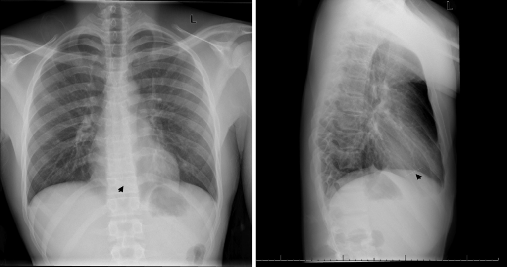
The chest x-ray (CXR) and Computed tomography (CT) scan helped paint the complete picture for the doctors. CXR revealed a linear density in the mid anterior chest, concerning for foreign body. The CT scan helped identify the foreign body as a linear metallic foreign object, 3.5 cm in length, partially lodged in the right ventricle of the teen’s heart.
The patient initially denied any foreign body ingestion however, in a later interview he admitted that he stitches his own clothes and is sometimes known to hold sewing pins in his mouth so as to not misplace them. However, he had never knowingly ingested a sewing pin.
Fortunately, the sewing pin was successfully removed from the right ventricle and the patient went on to recover without any complications.
This is believed to be the first case of an intracardiac needle as a result of occult ingestion. According to Dr. Bonnie Mathews, lead author of the study, the pin may have migrated directly from the stomach into the heart, or from along the gastrointestinal tract.
The findings of the study demonstrate the need to consider foreign body ingestion as a differential diagnosis for perimyocarditis especially when the typical symptoms are absent. As well as, in high-risk individuals such as the pediatric population, psychiatric patients, I/V drug addicts or someone with recent medical instrumentation.
As there can be devastating consequences of ingesting sharp foreign objects, it is vital that doctors consider the removal of gastric foreign bodies to prevent complications due to migration as in the case of this 17-year-old.
Reference:
Mathews, B., Chen, C., & Fahey, M. (2020). Occult Ingested Foreign Body: An Unusual Cause of Perimyocarditis. The Journal of Emergency Medicine.doi:10.1016/j.jemermed.2020.06.036


