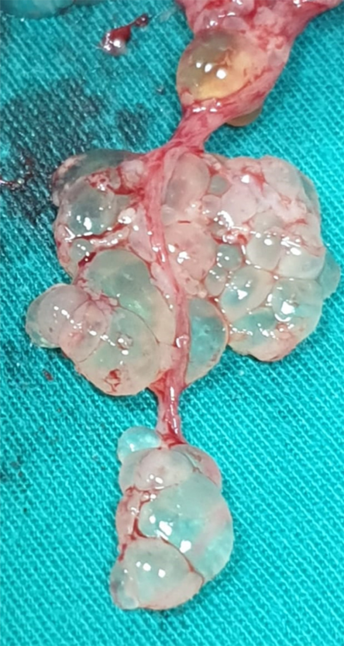
Invasive mole, a manifestation of gestational trophoblastic disease
Gestational trophoblastic disease (GTD) is defined as a spectrum that consists of premalignant and malignant conditions. Currently, there is no literature on how fast the invasive moles can develop from choroideremia. If the condition persists after initial treatment, with elevated beta-human chorionic gonadotropin, it is known as post-molar gestational trophoblastic neoplasia (pGTN). Currently, there is no detailed information on how fast these moles develop from CMH. However, the risk of developing it is the highest within the first 12 months. Similarly, most cases present within the first 6 months. In this case, the patient presented with a uterus size greater than the gestational age and was diagnosed with an invasive mole.
Case study
This article highlights a similar case, in which a 46-year-old primigravida woman presented to the outpatient clinic with a uterus size greater than the gestational date. She had last menstruated 8 weeks ago and her pregnancy test was positive. The patient did not have a history of a miscarriage or abortion. Vital signs were normal. Physical examination was consistent with a large uterus, which is normally the case in the 16th week of pregnancy. Further laboratory tests showed an elevated β-hCG serum level (>300 000 mIU/mL), whereas other parameters were normal.
For further evaluation doctors advised an ultrasound that showed a hydatidiform mole
Treatment included suction, evacuation and curettage. Reevaluation after the procedure showed that the mole was completely removed. Samples were sent for macroscopic and microscopic evaluation to the pathology anatomy lab. Findings showed a complete hydatidiform mole without any signs of malignancy.
The patient was referred for a follow-up on the 7th, 14th and 21st days after the procedure. However, the patient did not return for a follow-up on the 14th day because of a significant reduction in β-hCG serum levels. On the 22nd day, the patient presented to the emergency room with severe vaginal bleeding. Doctors advised an ultrasound that showed fluid in the uterine cavity. The patient’s haemoglobin level had fallen to 6 mg/dl, whereas her β-hCG serum level increased to 53 969 mIU/ mL.
Based on these findings, doctors diagnosed the patient with gestational trophoblastic disease (GTD). X-rays findings did not show any signs of distant metastases. After discussing with the patient, the patient opted for a hysterectomy. 2 days later the hysterectomy was performed and the specimen was sent for evaluation. The findings were consistent with the diagnosis of an invasive mole.
Source: American Journal of Case Reports


