Hydatid cyst of the foot, initially suspected to be a callus
Cystic echinocccosis (CE), also referred to as hydatid cyst is caused by infection with Echinococcus granulosus, a long tapeworm found in dogs, sheep, cattle, goats and fish, in its larval or cyst stage. Although the infection is asymptomatic in most cases, it can cause slowly enlarging cysts in the liver, lungs and other organs. The cysts are often neglected and go unnoticed for several years. This article describes the case of hydatid cyst in the plantar surface of the patient’s foot. The cyst is rarely located in soft tissues, therefore, making this an extremely rare case.
The 22-year-old Somali patient was referred to the surgical clinic of Djibouti Medical Center in March, 2019. The patient complained of progressive swelling associated with pain in the left plantar side of the foot. There were no signs of abdominal pain or discomfort, chest pain or cough. According to the patient, the swelling appeared a year before his presentation to the clinic.
The patient’s medical history did not reveal any incidence of trauma to the site
He had no difficulty walking, discharge from swelling or lesions on other sides. The 22-year-old was a college student living with his parents and had no history of any medical problems. Similarly, according to his family history, there were no medical illnesses running in the family. He had no history of tobacco smoking or substance abuse. And he had never consumed alcohol.
On examination, the patient’s blood pressure was 120/70 mmHg, respiratory rate was 18 per minute, pulse rate was 74 beats per minute and temperature was 36.0°C axillary. The rest of the physical examination showed no significant findings. There were no neurological abnormalities noted, either. Examination of the patient’s foot was consistent with an oval-shaped, well-circumscribed lump, measuring 2.5 cm wide in diameter. The lesion was non-pulsatile and located over the left foot in the mid-planar. The lump was fluctuant and tender to touch with change in colour of the overlying skin.
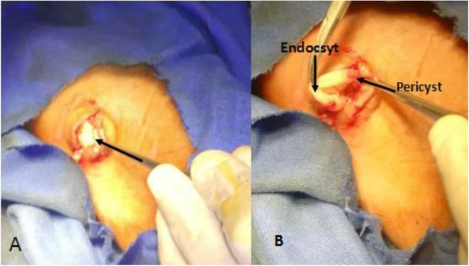
Doctors initially suspected for the lesion to be a callus and prepared the patient for a minor surgical operation for removal of the mass. However, dissection of the area revealed an endocyst and pericyst, consistent with the diagnosis of a hydatid cyst very clearly. Following the surgery the patient was prescribed with diclofenac 50 mg twice a day for 5 days. The patient was called back for a follow-up after a week which showed satisfactory wound healing.
References
Hydatid cyst of the foot: a case report https://jmedicalcasereports.biomedcentral.com/articles/10.1186/s13256-019-2337-8


