Case Presentation
A 36-year-old nulliparous woman from Nigeria presented to a clinic with the complain of painless abdominal mass for one year. According to the patient, the mass had increased in size since it first appeared
History and Examination
On abdominal and pelvic examination, the doctors observed a mass that was consistent with the size of a 36-week gravid uterus. It seemed to be originating from the pelvis and was not tethered to any overlying structures. The rest of the examination was unremarkable.
Her past gynaecological history was non-significant. However, she was on anti-hypertensive medication amlodipine, 10 mg per day. Moreover, her surgical history consisted of an open myomectomy that was done three years back due to fibroids. She also had a pelvic ultrasound done prior to presentation, which was suggestive for adenomyosis.
Investigations and Work-up
The doctors sent a repeat ultrasound scan, along with baselines, for the assessment of the abdominopelvic mass found on examination. This was done in order to prepare the patient’s pre-operative workup. The lab reports showed a packed cell volume (PCV) of 36%, and fasting blood sugar of 80 mg/dl. The abdominopelvic ultrasound showed an anteverted, and bulky uterus with dimensions of 283 × 156 mm. The endometrium was 4 mm thick, along with myometrial diffuse enlargement. The rest of the findings were non-significant; therefore, the doctors made a diagnosis of adenomyosis. CT and MRI could not be performed due to unavailability of these investigation facilities in the clinic.
Surgery: Removal of the Abdomino-pelvic mass
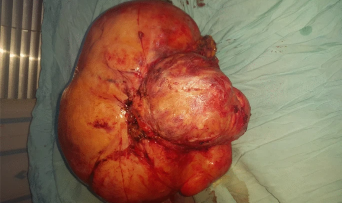
The doctors performed an explorative laparotomy to manage adenomyosis and to remove the mass in the uterus. During the laparotomy the doctors found a firm tumor of 40 cm diameter, weighing 6.5 kg. The tumor was adherent to the omentum, with enlarged omentum vessels. The ovaries, fallopian tubes and the retroperitoneum were not involved. However, the tumor extended towards the sub-umbilical area of the abdominal wall. The doctors excised the tumour along with wide free margins. Moreover, they also performed a partial omentectomy. The excised mass was sent for histopathology.
The woman was stable postoperatively and no complications were observed. The doctors discharged her on the 7th post-operative day.
Diagnosis: Desmoid Tumor
The histopathology report of the excised tumor revealed a desmoid-type fibromatosis. The doctors decided to follow up with the patient for the subsequent months. Ten months after the patient’s surgery, she presented with an incisional hernia. Thus, the doctors decided to perform surgery to manage the hernia. The surgery was uneventful, with no post-operative complications.
Prospect and Future Implications: Desmoid Tumor
This case highlights the need for timely intervention for abdominopelvic masses, especially in areas where facilities such as MRI and CT scan are not available. A delay in diagnosis may lead to delayed surgery and thus growth of tumor, in addition to other complications related to the tumor. Therefore, when encountering an abdominopelvic mass in females should always raise the suspicion of any of these masses or tumors : leiomyoma, desmoid tumors, ovarian cyst, and ovarian malignant tumor. In this case, the female was offered hysterectomy at a clinic after the first ultrasound scan which revealed the abdominopelvic mass, which may have lead her to consult a local clinic. This eventually lead to an explorative laparotomy, which aided in timely management and diagnosis of the tumor.
Desmoid Tumor: Overview
Desmoid tumours are benign tumours that originate from the fascia and aponeurosis of a muscle. They can grow in any area of the body, and can be classified into benign and aggressive tumors. The former is not associated with FAP, whereas, the latter is associated with FAP.
They can be asymptomatic and may even present as pain, local infiltrating mass, loss of function in the affected area, nausea and cramps.
The risk factors the predispose young women to the development of desmoid tumors are young age, pregnancy, muscle injury, history of colectomy, contraceptive use and a family history of FAP. Moreover, an association between previous abdominopelvic surgery and the development of desmoid tumor is seen due to the risk of development of the tumor on previous scars in this region.
The investigations that are used to diagnose desmoid tumors are radiological scans such as CT, Ultrasound and MR. Definitive diagnosis, however, is always made on histopathology.
The management of desmoid tumors varies due to the unpredictable nature of the growth of the tumor. This is because the pathophysiology of the tumor is poorly understood. Treatment depends on the stage of the tumours ( based on growth, size and presence of symptoms). Stages I, II and III can be managed with a variety of treatment modalities such as as non-steroidal anti-inflammatory drugs (NSAIDs), selective tyrosine kinase inhibitor, anti-estrogens, chemotherapy, and surgical resection. However, Stage IV tumors, tend to show aggressive growth along with aggressive and early presentation of symptoms. Moreover, the risk of complications and early death is drastically increased. Therefore, medical and surgical management is usually impossible.


