
Case Presentation
A 51-year-old man from with no significant medical history presented with acute low back pain and pain radiating to the right leg. He had no obvious trauma or any other significant events. He was employed, had no significant family history, and did not smoke or consume alcohol. Doctors administered a sacral epidural block but was referred to this department when it did not help with the pain. Neurological examination showed a positive straight leg elevation test 80° to the right. Manual muscle examination revealed weakness of the right leg (tibialis anterior, 3/5; extensor hallucis, 3/5; flexor hallucis longus, 4/5; and triceps surae, 4/5). The right patellar tendon reflex was normal and the right Achilles tendon reflex was slightly impaired. Doctors noted hypoesthesia at the right L5 dermatome. Neurologically, these results indicate L5 nerve root dysfunction.
Investigations
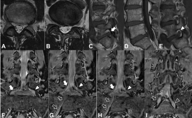
Magnetic resonance imaging (MRI) did not show a herniated disc. However, doctors saw a right combined lumbosacral nerve root on T2-weighted imaging with no foraminal stenosis on T1-weighted imaging. The position of the right L5 nerve root in the intervertebral space was significantly lower than that of the left L5 nerve root, and the right L5 nerve root joined the common trunk with the S1 nerve root. The right S1 nerve root branched closer than the left S1 nerve root. Doctors diagnosed him with Conjoined lumbosacral nerve root (CNR). He received pain and anti-inflammatory medications and a selective nerve root block.
Surgery: Combined Lumbosacral Nerve Root
After a month, however, the pain did not subside and we performed a partial laminectomy. Laboratory evaluations showed no evidence of an inflammatory reaction (C-reactive protein 0.09 mg/L. White blood cell count 4.0 × 10⁹/L, and platelet count 212 × 109/L). Laboratory evaluation of liver and kidney function showed no abnormalities [aspartate aminotransferase (AST) 29 U/L, alanine aminotransferase (ALT) 39 U/L, alkaline phosphatase (ALP) 170 U/L. Blood urea nitrogen (BUN) 17.8 mg/ dl and creatinine 0.83 mg/dL]. Urine, serological and microbiological analyzes showed no abnormalities.
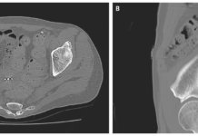
His body temperature on admission was 36.7°C, pulse 56 beats per minute, and blood pressure 125/69 mm/Hg. Intraoperatively, doctors discovered that the right L5 root originated from the caudal plane of the L5 pedicle and joined the right S1 nerve root. They dismantled the CNR by facet joint resection. Postoperative three-dimensional computed tomography (3D-CT) showed a successful partial laminectomy. They partially removed the right facet joints L4-5 and L5-S. Immediately after the operation, the symptoms completely disappeared.
Recurrence of Symptoms
But the same symptoms recurred 7 years after the operation. However, MRI showed no disc herniation on T2-weighted images, but T1-weighted images showed superior L5-S foramen stenosis. The right L5 nerve root (related nerve root) was compressed due to right foraminal stenosis caused by L5-S disc degeneration. A neurological examination showed a negative result of the test of lifting the right leg. Hand muscle examination revealed weakness of the right leg (tibialis anterior, 3/5; extensor hallucis, 3/5). Right patellar and right Achilles tendon reflexes were normal. Doctors noted hypoesthesia at the right L5 dermatome. Neurologically, these results indicate L5 nerve root dysfunction.
Diagnosis
She was diagnosed with right L5-S stenosis, received pain and anti-inflammatory medications, and underwent a selective sacral epidural nerve root block. However, her pain did not improve after 3 months and we performed L5-S transforaminal lumbar interbody fusion (TLIF). Laboratory evaluations showed no evidence of an inflammatory reaction (C-reactive protein 0.10 mg/l, white blood cell count 5.5 × 10⁹/l, and platelet count 204 × 109/l). Laboratory evaluations of liver and kidney function were unremarkable (AST 28 U/L, ALT 38 U/L, ALP 176 U/L, BUN 18.4 mg/dL, and creatinine 0.81 mg/dL).
Urine, serological and microbiological analyzes showed no abnormalities. At the second admission, his body temperature was 36.5 °C, pulse 53 beats per minute, and blood pressure 106/72 mm/Hg. In the second operation, a right L5-S fasciectomy revealed that the right CNR crossed the L5-S disc space and there was a significant swelling of the right CNR, which made it immobile. To perform TLIF, the right CNR must be safely retracted medially, the intervertebral disc removed, and an 11-mm-high, 10-mm-wide drill placed into the L5-S disc space. Doctors decided that if they create this space with surgical techniques, unfortunately there is a high risk of excessive traction compromise and superior CNR. Therefore, they rejected insertion from the left side and performed TLIF from the contralateral side. Immediately after the operation, the symptoms completely disappeared.
Combined Lumbosacral Nerve Root
Conjoined lumbosacral nerve root (CNR) is an embryological nerve root anomaly that primarily affects the lumbosacral region. Abnormal roots are primarily bidirectional, interconnected structures that arise from a large area of the dura. Because of their size and attachment to surrounding structures, these roots are uniquely vulnerable to trauma. The effects of compression and entrapment are enhanced in the lateral fossa joints, where developmental changes and disc herniation reduce the available free space. Adequate root exposure may be necessary in patients with Conjoined lumbosacral nerve root (CNR) to prevent continuous compression and reduce traction. Therefore, hemilaminectomy must be performed in such a way that the intervertebral space or lateral fossa is sufficiently open to avoid adverse changes in stability. Moreover, to ensure adequate mobility of the lumbosacral spine.