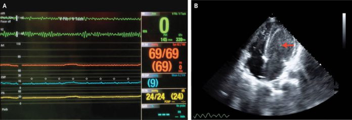
A 50-year old woman presented to the emergency department with a 1-week history of weight gain, fatigue and dyspnea. She also had a history of bicaval cardiac transplantation. A bicaval heart transplant is a simple surgical technique in which the right and left atria of the donor is incised and anastomosed to the posterior wall of the atria of the recipient’s heart. Until the 1990s, this was a standard heart transplant surgical technique.
A transthoracic echocardiogram of the heart showed a reduced systolic function in the left ventricle. In addition, acute cellular rejection of the transplanted heart was confirmed via an endomyocardial biopsy. The hemodynamic status of the patient also deteriorated, moreover, prompted an initiation of venoarterial extracorporeal membrane oxygenation.
Further investigation revealed the development of a sustained ventricular fibrillation and loss of right and left ventricular contractility as shown by a non-pulsatile pulmonary arterial waveform, in addition to a systemic arterial waveform. However, the central venous waveform was observed as regular pulsatile.
The echocardiography of the patient was repeated which confirmed a left ventricular thrombus with near ventricular standstill and the right atrial contractility, corresponding to the central venous waveform was preserved with tricuspid valve opening.
The findings, including concomitant mitral valve opening were consistent with atrioventricular dissociation during ventricular fibrillation. Moreover, premature closure of atrioventricular valves was also observed right after atrial contraction. Atrioventricular dissociation is a condition in which the atria and ventricles activate independent of each other. It presents with symptoms including fatigue, malaise, exertional dyspnea, palpitations, throbbing neck sensations and lightheadedness.
The patient suffered from multi-organ failure and passed away a week after presentation.
References
Salik, J. R., & Thomas, S. S. (2020). Atrioventricular Dissociation during Ventricular Fibrillation. New England Journal of Medicine, 382(23), e84.


