According to the World Health Organization (WHO), cancers accounted for an estimated 9.6 million deaths in 2018. Early detection of cancers and adequate treatment of patients can play a huge role in reducing the cancer burden. However, early detection can be a real challenge without good imaging contrast agents.
Contrast agents are usually injected into patients during imaging techniques such as MRI and CT scans to help detect tumors and, to help surgeons identify accurate tumor margins. Currently, only three optical imaging contrast agents have been approved by the FDA for use in humans.
Scientists Develop New Imaging Contrast Agents
In the hopes to improve patient outcomes, researchers at the University of Southern California (USC) have developed new imaging contrast agents using common food dyes and tattoo ink. The research was conducted at USC’s Viteri Department of Biomedical Engineering and led by Cristina Zavaleta.
For their research, Zavaleta and her team chose FDA approved food dyes such as those found in cosmetics and candies and, evaluated their optical properties.
Tattoo Ink Detects Cancer
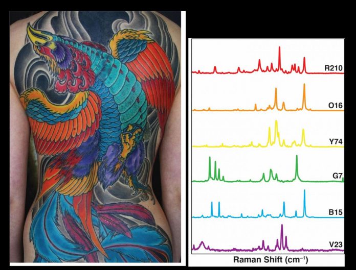
The idea to use tattoo ink as a contrast agent came to Zavaleta when she was attending an animation class at a Pixar studio. The bright watercolors around her made her wonder whether they could have optical properties. Which led her to a tattoo artist in San Francisco called Adam Sky.
When she ran the tattoo ink through a Raman Scanner, she found each color possessed a unique spectral fingerprint that could be used to barcode their nanoparticles.
Nanoparticles Illuminate Cancers
The food dyes and tattoo ink were then paired with nanoparticles, which illuminated the cancer cells. By allowing doctors to differentiate between cancerous and non-cancerous cells, illuminated nanoparticles could provide a more sensitive imaging contrast.
If you encapsulate a bunch of dyes in a nano-particle, you’re going to be able to see it better because it is going to be brighter. It’s like using a packet of dyes rather than just one single dye
Cristina Zavaleta
The new contrast agents were found to have better absorption, fluorescence, and Raman scattering properties, making them the ideal ‘optical inks’ to be attached to nanoparticles. Nanoparticles, developed by the team to carry these optical inks, were believed to be small enough to permeate the tumor cells and stay retained in them longer than the commonly used dyes.
However, there are a few challenges involved with using nanoparticles for imaging. They can have potential toxicity effects due to their prolonged retention in organs such as the liver and spleen. Therefore, it is important to consider biodegradable and biocompatible nanoparticles.
The team of scientists believes that the set of dyes they have discovered, if better developed, can lead to great improvement in patient outcomes and reduce mortality.
Reference:
Helen R. Salinas et al, A colorful approach towards developing new nano-based imaging contrast agents for improved cancer detection, Biomaterials Science (2020). DOI: 10.1039/D0BM01099E


