
Doctors detect bilateral myxomas in this 15-year-old through thoracic CT and echocardiography!
A 15-year-old female presented to the hospital with a complaint of cardiopulmonary distress. She reported that the disturbances began nearly 20 days ago. On her current presentation, she was tachypneic and tachycardic. Her temperature was elevated too. Her physical examination revealed pale conjunctiva and oedema of the face. Doctors heard systolic murmurs and a very loud apex beat. They also noticed lower limb oedema and an enlarged liver in the patient.
Masses in the Ventricles
Laboratory investigations of the patient revealed decreased albumin and elevated brain natriuretic peptide levels. Doctors next obtained a radiograph of the patient’s thorax which revealed mild pneumonia with pleural effusion on the right side. On a transthoracic ECHO, the doctors found multiple ventricular masses. One mass was present in the right ventricle. It prolapsed during each systole into the right atrium through the tricuspid valve. Another was present in the right atrium itself. Moreover, doctors also found a mass in the left ventricle near its outflow tract.
Bilateral Myxomas Confirmed!
The masses together with other lab findings suggested bilateral myxomas. However, doctors also put vegetations and thrombi in their differentials. They performed a thoracic CT that confirmed the masses and also revealed the presence of mild pericardial effusion. A fluorodeoxyglucose test suggested the masses to be benign tumours.
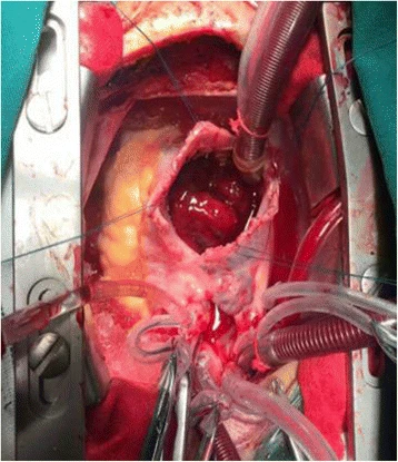
Moreover, they kept the patient on pre-op protocols for 2 weeks to improve her nutritional and cardiopulmonary status after which they performed the surgery. They operated upon the patient under general anaesthesia and approached the heart via a median sternotomy. After carefully identifying the tumours in both ventricles, they removed them with 10mm margins. For a right ventricular tumour that prolapsed into the right atrium, they approached it through the tricuspid valve. They then repaired the valve after removing the tumour.
Histopathological reports confirmed bilateral myxomas. Doctors kept the patient for a ten-year follow-up. Fortunately, the patient remained healthy and did not show any signs of recurrence.
Cardiac Myxomas: An Insight
Cardiac myxomas are one of the most common primary tumours of the heart. Their frequency peaks in females and most commonly, they occur in the left atrium. However, they can develop in any of the four chambers of the heart. Most myxomas have a stalk and a base and their surface can show villous morphology. Moreover, they can also be globular or jelly-like in gross appearance.
Histologically, myxomas consist of myxoid connective tissue in which the cells take up hematoxylin stain. The reason lies in the fact that these cells are rich in mucopolysaccharide content. The cells range from stellate to fusiform shapes and stain positive for a number of immunohistochemical markers such as S-100, vimentin and CD56 etc.
Signs and Symptoms
Patients with myxomas develop symptoms primarily due to obstruction of blood flow inside the cardiac chambers. They present with symptoms such as anaemia, cardiovascular distress, fatigue and weight loss. Sometimes, they also develop a cardiac rumble. However, many patients remain asymptomatic too which ultimately leads to a delayed diagnosis. Anyhow, for all patients with suspected myxomas, transthoracic ECHO remains the test of choice. It provides detailed visualization of heart chambers and thus helps in identifying any tumours.
Treatment and Prognosis: A Double-Edged Sword
Myxomas once identified need treatment through surgery. However, removing these tumours comes with its own risks and complications. Surgical removal of these cardiac masses especially in the right ventricle can lead to intraoperative damage to cardiac valves and consequent valvular and cardiac insufficiency. Mortality during the procedure comes out to be two to five percent. On the other hand, if the diagnosis is not followed by a prompt surgery, the risk for embolism increases which can result in sudden death. Thus, it’s a double-edged sword and while doctors have to opt for immediate surgery, they have to do it under great caution to avoid any intraoperative complications.
Our patient developed bilateral myxomas which are even more difficult to treat. Moreover, the involvement of the tricuspid valve and a prolapsing cardiac mass made this case rarest of rare. Anyhow, the doctors not only successfully removed the masses but also skillfully repaired the tricuspid valve with everything turning out well in the end.


