Case Report
Tumor deposits (TDs) are tiny, irregular tumor clusters that exist in adipose tissue separate from the underlying tumor. They have been reported in a variety of tumors, including breast carcinomas. Multiple studies have revealed TDs as independent indicators of early recurrence and poor prognosis in gastric and colon malignancies.
Their therapeutic role has also proven in colorectal cancer, where TDs alter the N classification and, as a result, can determine the kind and duration of adjuvant therapy. Researchers have detected extra nodal tumor deposits (ETDs) in 10% of breast cancer cases and have determined that they can predict the development of four or more additional positive nonsentinel lymph node metastases.
Some scientists have proposed that this new entity to include in breast cancer’s tumor, node, and metastasis (TNM) staging. The current version of the TNM classification of breast cancer does not include TDs in the N staging, which might lead to an underestimate of the tumor stage and, as a result, inadequate treatment. The study will focus on TDs in breast cancer and their possible impact on prognosis and TNM categorization.
Case Presentation
A 58-year-old Tunisian female patient with no personal or family medical history reported a palpable lump in her left breast. Physical examination indicated a hard, poorly defined lump in the outer quadrants of the left breast, along with skin alterations suggestive of “peau d’orange.” The bulk was 2 centimeters in diameter. No palpable axillary lymph nodes were found. Clinically, the nodule was designated as cT4bN0Mx.
Imaging and Investigation
Breast ultrasonography and mammography both identified the lesion as American College of Radiology category 5 (ACR5). A core needle biopsy verified the presence of invasive breast cancer of no special kind, Scarff-Bloom-Richardson (SBR) grade III, luminal B, and HER2-positive subtype. At the time of diagnosis, further imaging revealed no distant metastases.
Treatment
The patient underwent four cycles of neoadjuvant chemotherapy, which consisted of epirubicin-cyclophosphamide followed by paclitaxel, over the course of six months. The treament included chemotherapy along with hormonal treatment and radiotherapy . Following chemotherapy, ultrasonography and mammography confirmed a full response. Later, the patient had a left mastectomy with ipsilateral axillary lymph node dissection (ALND).
The mastectomy specimen showed a fibrous, white plaque of 15 cm×9 cm×4 cm with necrosis. It was in the outer quadrants, away from the surgical margins. The axillary dissection specimen was 10 cm × 8 cm × 2 cm and had 28 nodules ranging in size from 0.3 cm to 1 cm.
Histologic Finding
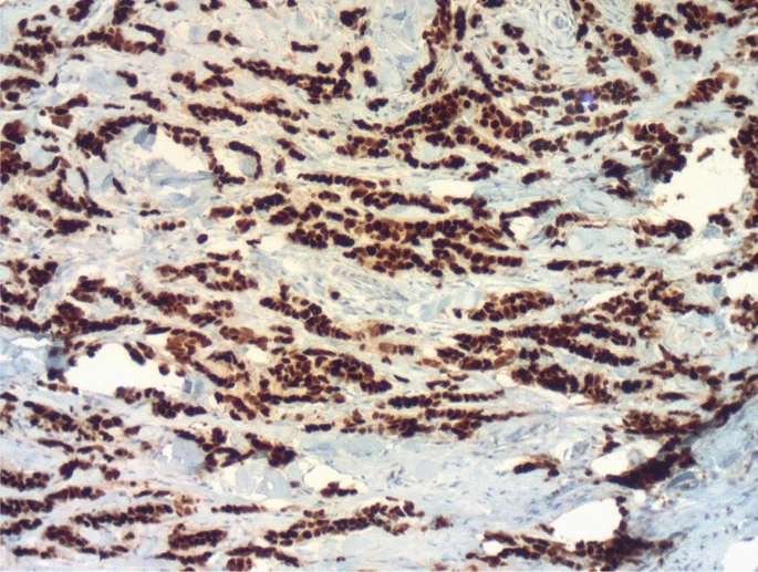
Extensive sampling of the fibrous area revealed extensive scar fibrosis, along with elastosis and a few inflammatory cells, indicating a partial response to chemotherapy. More over half of the tumor was still alive. The tumor cells were medium-to-large in size, with eosinophilic or pale cytoplasm and big nucleoli. A vascular embolism was discovered, but no perineural invasion or Paget’s disease were observed. All surgical margins were disease-free.
Five of the 28 recovered nodules were metastatic lymph nodes, with two exhibiting extra capsular expansion. The remaining 14 nodules were classified as TDs. They were tumor clusters that existed independently, with no lymphocytic peripheral rim or remnant vascular or neural walls. The tumor staged as ypT1aN2aM0. The patient continued to undergo hormone therapy along with trastuzumab. The follow-up revealed no issues. The patient remains in good clinical condition. There is no evidence of a recurrence.
Discussion
In this case, we found 14 TDs and five lymph node metastases in the axillary area of a woman who had a mastectomy and axillary dissection for a high-grade luminal B HER2-positive invasive breast cancer. In this scenario, TDs were identified using the same criteria as for colorectal cancer (CRC). These criteria include the absence of a lymphocytic peripheral rim and capsule, as well as the lack of a remnant vascular wall or neural tissue within the tumor cluster. Although TDs were not included in the N classification, their presence and number were clearly reported in our final pathology report.
TDs are a relatively new type of breast cancer. There are very few research studies on TDs in breast malignancy. In actuality, it is a contentious entity, with recent articles and statistics. Their frequency ranged from 10% to 29%. However, TDs were first documented in colorectal adenocarcinomas, and thereafter in other carcinomas, including head and neck, pancreatic, biliary, stomach, and cholangiocarcinoma.
In this example, the residual tumor was assessed as greater than 50%. This partial response is a common finding in T4 tumors, with studies revealing that more than half of T4 tumors obtained partial response regardless of whether TDs were present or absent. However, data on the association between TDs and chemotherapeutic response are sparse and limited.


