Case report: carney complex
This article describes the case of an otherwise healthy, 54-year-old male patient who presented with an atypical presentation Carney complex. The patient complained of an intermittent and productive cough with a history of 2 years. In addition, his dyspnea had worsened over the past 2 to 3 months. He could barely walk a few feet without dyspnea. During this time, he also struggled with palpitations and intermittent lightheadedness. However, there were no signs of upper respiratory tract infection symptoms, fevers, chills or chest pain. He had no recent travel or smoking history.
On his previous visits to urgent care, he was diagnosed with bronchitis and pneumonia. For treatment, doctors prescribed antibiotics and steroids. However, the patient showed no signs of improvement despite treatment. For further evaluation, the patient was referred for a chest X-ray which showed an apparent retro-cardiac opacity. A CT scan was also done which showed an abnormal filling defect in the left atrium, extending into the left ventricle.
The patient was referred for cardiology evaluation
Physical examination showed that the patient was afebrile, with oxygen saturation 99% on breathing ambient air, blood pressure 136/81 mmHg and heart rate 94 beats per minute. There were no signs of respiratory distress, whereas cardiac exam showed a normal heart rate with regular rhythm. Lung examination was also unremarkable. However, skin examination showed multiple flesh-coloured skin tags around the circumference of the neck, anterior left ear, back and right upper cheek.
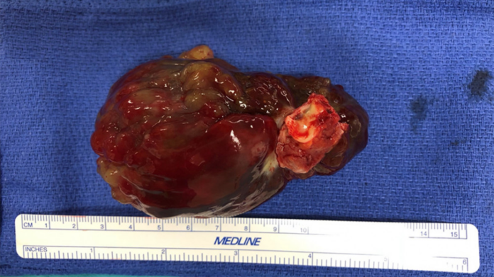
His previous medical history was also unremarkable. Although his family history was significant for diffuse skin tags in his father and an infant daughter who passed away of an unknown congenital heart disease. Doctors further advised a transthoracic echocardiogram which showed a left atrial mass measuring 9×4 cm with a mildly reduced ventricular ejection fraction. Transesophageal echocardiogram confirmed these findings and showed a large left atrial mass measuring 7.5×4.5 cm.
Based on these investigation findings, the patient was diagnosed with carney complex
Carney complex is a hereditary condition which is associated with spotty skin pigmentation and noncancerous myxomas (connective tissue tumours). It may also present with endocrine tumours. Symptoms typically develop in their early 20s. Heart myxomas and other heart problems are usually the first signs of carney complex.
The patient was referred for urgent surgical excision of the mass. The mass was further sent for histopathological analysis which confirmed the diagnosis of cardiac myxoma. Doctors discharged the patient with close follow-up.
Source: American Journal of Case Reports


