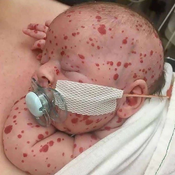
An interesting case of a child who presented with multiple hemangiomas on the skin. Hemangiomas are bright red birth marks that show up either in the first or second week of life or at birth. The hemangioma is made up of extra blood vessels and has the presentation of a rubbery bump in the skin. Hemangiomas commonly appear on the back, chest, scalp and face, however, can occur anywhere on the body. The hemangiomas in babies, known as infantile hemangioma fade over time, therefore, there is no treatment needed. Children who have this condition during infancy have visible traces of growth of the hemangiomas by the age of 10. Treatment needs to be considered if the hemangioma interferes with breathing, seeing or any other functions1.
A hemangioma most often
Depending on the size and location of the hemangioma, they do not usually cause any symptoms neither after nor during their formation. However, if they are in a sensitive area or grow large, they may cause some symptoms. Hemangiomas on the skin because of their deep red appearance are sometimes called strawberry hemangiomas. If hemangiomas are present inside the body, the symptoms are specific to the organs that are affected, for example, if the gastrointestinal tract or liver is affected, the symptoms include, a feeling of fullness, loss of appetite, abdominal discomfort, vomiting and nausea.
The diagnosis of hemangiomas in children is usually made with physical examination and visual inspection; hemangiomas in organs may only be spotted during an imaging test, CT scan, ultrasound or MRI. Moreover, they are usually detected by chance in some circumstances. Doctors monitor hemangiomas during routine checkups, the doctor needs to be contacted if the hemangioma looks sore, infected or bleeds2. A hemangioma is made up of a group of extra blood vessels into a dense clump. However, what causes the vessels to clump is not known.
Hemangiomas occur in babies who are white, female and born prematurely. If hemangiomas break down, a sore is developed, leading to bleeding, pain, infection or scarring. In addition, the hemangioma may interfere with a child’s hearing, breathing or vision, depending on where the hemangioma is situated, however, this is quite rare.
References
- Metry DW. Infantile hemangiomas: Epidemiology, pathogenesis, clinical features, and complications. https://www.uptodate.com/contents/search. Accessed Feb. 22, 2019.
- Metry DW. Infantile hemangiomas: Management. https://www.uptodate.com/contents/search. Accessed Feb. 22, 2019.


