This case report is of a 38-year-old Iranian non-smoker woman who appeared with a nonproductive cough. It started as a consequence of overusing antiseptic products to prevent coronavirus infection. However, her symptoms began quite before the COVID-19 virus. However, exposure to fragrances, powerful aromas, and spices exacerbated the sporadic acute cough, which lasted for several days. Before asking for help from a doctor, the patient’s symptoms continued for around three months. Moreover, she experimented on a variety of antitussive drugs. However, she did not get the best results. She had no unusual medical conditions other than allergic rhinitis. There was no past medical history of heart illness, lung disease, chronic sinusitis, TB, or gastroesophageal reflux disease.
Physical Examination
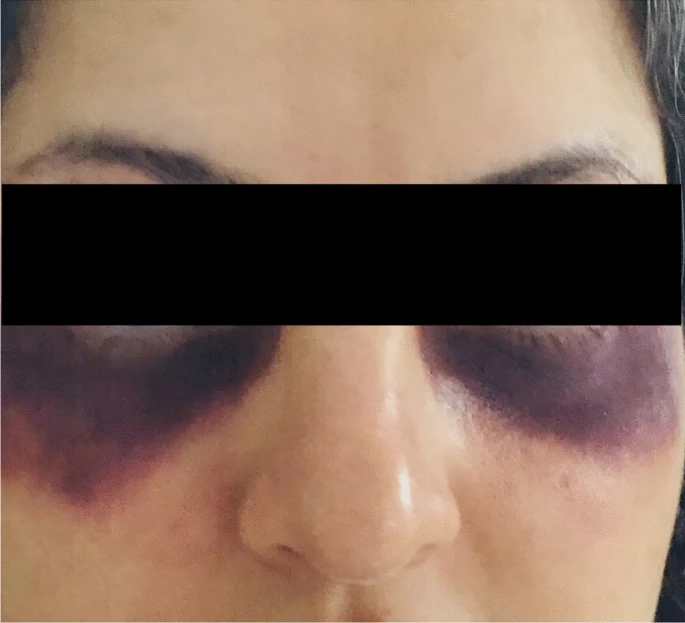
Moreover, since she was a housewife, she did not have any exposure to harmful toxins. However, for the previous two years, she had misused antiseptic products to ward off the coronavirus illness. Doctors did a physical examination, which revealed no unusual audible sounds, such as wheezing or rhonchi, and was otherwise normal. In addition, a reddish-purple rash on and around the eyelids resembling a heliotrope rash was also seen. The likeliness of the development was due to her persistent cough.
The pulmonary function test results were initially within normal ranges. However, after using a bronchodilator, a significant rise from baseline was seen in FEV1. Both the chest x-ray and the methacholine challenge test were normal. Hyperinflation and tree-in-bud opacities were seen on the chest HRCT.
Treatment
The patient was hospitalised and the doctors did a thorough diagnostic evaluation. Bacteriology and sensitivity tests on sputum produced negative results for several infections, even Mycobacterium tuberculosis. Doctors also ordered a CBC, which revealed mild hypochromic microcytic anaemia. Vitamin D3 levels were only marginally deficient and both cANCA and pANCA tests were negative. PT, PTT, and INR were also all within acceptable ranges.
SARS-CoV2 PCR came out empty. Cough-variant asthma was determined to be the most likely diagnosis after ruling out the other aforementioned potential causes of chronic dry cough. Doctors started an empirical asthma trial therapy was started for the patient. However, the outcomes were very unusual and surprising for the doctors. After receiving treatment for two weeks, there was a clear correlation between the disappearance of the heliotrope-like lesion over the eyes. In addition, a significant reduction in the cough was also seen. Furthermore, since the start of the therapy, the patient has been under strict observation continuously. In addition, she did not exhibit a return of the indications and symptoms either.
Discussion
Because of its diverse phenotypes and lack of a single diagnostic test to confirm or rule it out, asthma is a clinical diagnosis. The term “cough-variant asthma” refers to an asthmatic phenotype where the cough is the only outward sign. Spirometry examination with 12% and 200 ml improvement in FEV1 from baseline following bronchodilator challenge is the first-line method for diagnosing asthma. However, regarding the methacholine bronchial provocation test challenge. There is no general agreement, though. While some facilities conduct this test as a critical component of the work-up. It is not verifiable as a significant diagnostic test to evaluate the patient’s condition by others. Since asthmatic airway obstruction is variable. It may be difficult to establish objective evidence to support the diagnosis.
Lack of Testing for Asthma
Even if bronchial provocation and spirometry testing are available, a normal result does not rule out asthma. As a result, there is no gold-standard test for diagnosing asthma and related manifestations. Each patient’s test results must be uniquely evaluated. The bronchial wall thickening, areas of consolidation, bronchiectasis, and the tree-in-bud pattern are among the imaging findings of hazardous inhalation in HRCT.
Even though the pre-bronchodilator baseline measurements were completely normal. The patient’s FEV1 after bronchodilator medication showed increases from baseline. Methacholine challenge testing, however, came out negative.
The sole indications of hazardous inhalation on HRCT imaging were hyperinflation and tree-in-bud opacities. The patient also responded dramatically to the treatment. The eye darkening vanished as a result of the efficient therapy after admission and the beginning of appropriate asthma medication. Only in clinical situations is it advised to treat asthma before assessing reversibility with spirometry, according to the literature and current recommendations. Even though normal baseline measurements were reported, our patient’s pulmonary function test revealed reversibility, and her clinical situation was deemed critical.
When the patient was first admitted, the doctors carefully reviewed her medical history for any indications of possible exposure to dangerous airborne substances. Since she did not mention any exposures other than excessive use of the antiseptic solution. Hence, the doctors assumed that the antiseptic solution was the cause of the patient’s airway hyperreactivity. Moreover, she also avoided crowded places and strictly imposed social distancing. The following suggested that she had been exposed to indoor allergens. Therefore, antiseptic solutions were identified as the causative culprit and strongly recommended avoiding them while receiving medical treatment for asthma. This approach helped control the disease and reduce its symptoms.
Conclusion
Asthma is commonly assumed to be one of the diagnoses due to the various phenotypes and causes of cough. A thorough history, physical examination, objective lung testing, and potential therapy trials are all necessary for identifying individuals because asthma is a clinical diagnosis.


