
· Multiple gunshots were fired at a moving car.
· The car was carrying a 17-year-old young male.
· The bullet was reflected by the Nuss bar that was surgically inserted behind the patient’s sternum 7 months back
· The patient was saved by the Nuss bar and only suffered from minor flesh wounds.
· Penetrating wound injury was prevented by the Nuss procedure done to correct pectus excavatum 7 months prior to the gunshot incident.
A 17-year-old male presented to the emergency department with a gunshot wound to the left chest wall. The patient was in a moving car, which was shot repeatedly form an unidentified firearm. The car was hit by multiple bullets, but the patient was stable at the scene. He was taken to the nearby emergency department.
The patient had a history of pectus excavatum for which a Nuss procedure was performed seven months back. Nuss procedure is the preferred surgical procedure to correct pectus excavatum. One to three curved metal bars are inserted and left in situ behind the sternum to push the sternum into the normal curvature. The procedure is minimally invasive, and the bars are left inside for up to 3 years before being removed.
When the patient reached the emergency department, he was complaining of pain and chest wall tenderness on the left side of the thorax. There were no other complaints or symptoms.
Inspection: The patient was a thin and tall young male sitting with minimal discomfort with a shallow penetrating wound to the left chest.
Palpation: On palpation, the wound was tender.
Investigations:
The results of the complete blood count, metabolic profile, and serum troponin were normal.
Electrocardiogram:
Normal sinus rhythm with slight ST elevations in leads II, III, and aVF, with non-specific T wave inversion in V2, was seen on the electrocardiogram (ECG).
Radiography:
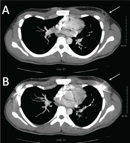
Two view chest radiographs revealed a single Nuss bar with bilateral stabilizing plates. There was no evidence of any bullet or its fragments. Any evidence of rib fracture, pleural effusion, or pneumothorax (Figure 1) was missing. A contrast-enhanced computed tomography scan of the chest was performed to confirm the findings of the chest Xray. The CT scan showed the induration of subcutaneous fat inferior and lateral to the left nipple, just anterior to the Nuss bar. There was no evidence of fracture, pleural effusion, pneumothorax, any vascular injury, or other intrathoracic injuries (Figure 2).
Considering the depth of the wound and the location of the wound, which was just anterior to the Nuss bar, it was noted that the bullet was most likely reflected by the Nuss bar, thereby preventing penetrating thoracic injury subsequently preventing any fatal consequences.
To rule out cardiac contusion, the patient was transferred to the trauma center where an echocardiogram was performed. ECHO showed a structurally normal heart without wall motion abnormalities. A repeat ECG was performed, which was showed no changes from the earlier performed ECG.
The patient was kept under observation overnight and then discharged.
References:
Pilegaard HK. Short Nuss bar procedure. Ann Cardiothorac Surg. 2016;5(5):513-518. DOI:10.21037/acs.2016.09.06
Serio F, McCague A (2017) Saved by the Bar: A Report of Bullet Deflection by Nuss Bar. Trauma Cases Rev 3:055. doi.org/10.23937/2469-5777/1510055


