Odontomas are mixed odontogenic tumours that consist of both epithelial and mesenchymal dental hard tissues. Moreover, single odontomas are the most common odontogenic tumours that are classified as either compound or complex.
Complex odontoma is a hamartoma in which enamel, dentin, cementum, and pulp are present. Furthermore, it is diagnosed mostly in children, adolescents, and young adults. Additionally, they usually occur as solitary jaw lesions. Nevertheless, if it is multiple odontoma, it is classified as a solitary jaw lesion, and it can also be present in all four quadrants of the jaw. However, since it is very rare, the clinical features are unknown.
This case report is about a 53-year-old woman and the first ever case of a patient of this age.
Case Report
She presented with a medical and dental history, which does not contribute to multiple odontomas. Furthermore, there was no significant medical history pointing towards it either. The patient’s chief complaint was swelling on the buccal side of her right horizontal body of the mandible. Additionally, the first permanent right molar was also displaced. The swelling was hard upon palpation and the adjacent premolar was mobile. However, no neck lymphadenopathy was seen.
The doctors did panoramic radiography and CT. They revealed two well-defined radiopaque lesions on the right mandible of the body. The lesion on the posterior region expanded the premolar and molar, externally and lingually towards the mandibular. Moreover, thinning of the lingual cortex and disruption of the buccal cortex were also seen.
Other than that, the remaining path of the IAN nerve canal seemed below the lesion. Moreover, both lesions were independent and intraosseous with the anterior one slightly disrupting and surrounding the mental foramen.
Based on the pathological and radiologic presentation, the diagnosis of provisional multiple odontoma was made. Moreover, the differential diagnosis included compound odontoma, ameloblastic fibro odontoma, osteoma, and cemento-ossifying fibroma.
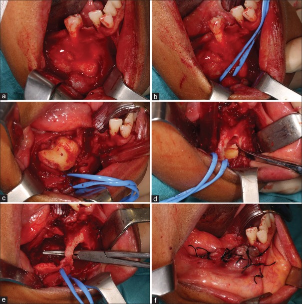
The doctors performed a conservative procedure of excision under general anaesthesia. They excised the posterior region. However, the lower second premolar could not be retained because of the lesion and periodontal status. Moreover, they separated the anterior lesion into two parts for enabling the excision. A thorough curettage was made after the excision. Additionally, the lesion was also sent for histopathology.
Since the lesion was rare for the lady’s age, histopathology was sent to two labs for analysis. Both labs confirmed the diagnosis of complex odontoma.
Recurrence and Follow-Up
When the patient followed up after fourteen months, no recurrence was seen. Moreover, the patient was asked to follow up regulary by the doctors.
Discussion
Numerous epidemiological studies have been made in the past based on the prevalence of odontogenic tumours. They are the most common tumours. However, multiple odontomas are very rare and the prevalence is unknown. Only two cases of multiple odontomas are identified out of which one of them is the case of a 53-year-old presented in this article. The female patient is the first eldest patient to be diagnosed and the tenth case described.
Generally, odontomas are asymptomatic. However, they appear at any age and are found mostly in the second decade. Moreover, they are not even gender related. The common symptoms include impaction, jaw swelling, and displacement of adjacent teeth. Additionally, malaligned teeth and pain are not very common.
In this case, the patient had a complaint of swelling in the jaw and pain, which started after the extraction of the lower right first molar. It started nine months before she visited the doctors with her concern.
Although the etiology of odontomas is still unclear, they are associated with traumas, inflammation, infection, and genetic causes. For example, Gardner’s syndrome, cleidocranial dysplasia, Hermann’s syndrome, and Pierre-Robin syndrome. Moreover, based on a study, the possible etiology of multiple odontomas is partial chromosomal duplication. It confers a gain of function of FGF3 and FGF4 genes. However, no family history was seen, and she was the first known patient of multiple odontoma in the family.
Complex odontomas are either spherical or ovoid in radiopacity. They have a fine radiating periphery, which is surrounded by a radiolucent zone. Moreover, it may broadly develop a complex odontoma. Moreover, a differential diagnosis of a compound odontoma and an osteoma may be impossible radiographically.
Conclusion
Multiple complex odontomas of the mandible are a challenge. Since most cases are close to vital anatomic structure. In this case the nerve was wrapped with a silicon tube after the lesion was dethatched, recognized, and localized. Hence, surgeons usually identify all the structures thoroughly to minimize the risks post-surgery. Moreover, no complication was seen after either.
Based on the extensive literature review and existing information, this is the oldest case report diagnosed.


