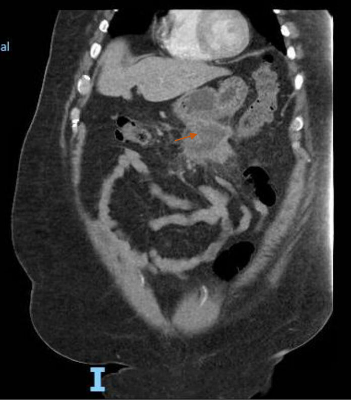
Idiopathic sclerosing mesenteritis is a very rare condition in which the adipose tissue goes through necrotic and fibrotic changes. It is common in Caucasian males between the age of fifty and seventy.
Case Study
A 60-year-old man presented to the hospital with widespread cramping pain in the abdomen for three weeks. The pain became worse a day before admission. However, it was non-radiating and nothing relieved or worsened the pain. The patient had a medical history of diabetes and hypertension.
There was no link of the pain with his diet and bowel movements. Although he did lose 30lbs weight over the past three months with a loss in his appetite. Moreover, he refused to have fever, chills, night sweats, nausea, vomiting, and constipation.
Diagnosis
A CT scan of the abdomen revealed a hypodense amorphous collection versus mass within the mesentery. Doctors suspected a necrotic tumour. Hence, they consulted the gastroenterology, general surgery, and interventional radiology team for a multifaceted approach.
After a second opinion from them, the patient underwent endoscopy (EGD), endoscopic ultrasound (EUS), colonoscopy and guided biopsy. Due to the size of the mass and proximity of the transverse colon.
EGD suggested an irregular hypoechoic mass in its longest axis. Moreover, from the distal position of the body, it showed to be outside the gastric echo layer. Doctors did the ultrasound-guided, FNB with four passes from the stomach’s distal body. However, the EUS was unremarkable. The common pancreatic duct and bile duct were not dilated. In addition, the colonoscopy revealed external and internal haemorrhoids.
The descending colon had an irregular, polypoid, granular appearing lesion surrounded by oedema, probably due to the erosive mesenteric mass or diverticulitis. Doctors did a cold forceps biopsy of the area. The biopsy results of the colonoscopy and EUS were non-specific.
Treatment
A diagnostic and therapeutic laparoscopic procedure was performed that revealed a large abscess cavity in the base of the mesentery, transverse colon, and loop of the small bowel. The doctors drained the abscess cavity and removed the inflamed mass in addition to the necrotic tissue. Histopathology confirmed the mass to be extensive fat necrosis. Furthermore, the nodules in the mesentery were significant for the diagnosis of sclerosing mesenteritis. His pain resolved and he was discharged with outpatient follow-up.
Sclresoing Mesenteritis – An Important Differential Diagnosis
Although sclerosing mesenteritis is a rare condition, it should be an important differential in patients with abdominal pain and mesenteric mass on imaging. It is a diagnosis of exclusion after ruling out other causes, for example, autoimmune diseases.


