In 2012, a mother brought her 4-month-old son to see his pediatrician about a stuffy nose. During the exam, the doctor noticed that the child’s head seemed to have grown larger than it should have in the two weeks since he last measured it.
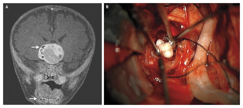
A contrasted MRI revealed that the baby had a large mass in his brain near the sella turcica. The scan also showed multiple mandibular teeth-like structures in the mass’ periphery. However, the doctors couldn’t decide what type of tumor it was without actually looking at it. They had to resect it.
Dr. Edward Ahn, a pediatric neurosurgeon at the Johns Hopkins Hospital led the risky surgery and approached through a right pterional incision. What they saw inside was quite surprising.
“We had to think twice,” he said. “We first thought they were flakes of calcium. When we looked at it closer, we were like, ‘Those really look like teeth.’ “
Histopathological testing then confirmed that those were indeed, fully formed teeth.
Classifying the tumor
The pathologists also confirmed that the resected mass was in fact, an adamantinomatous craniopharyngioma. This is a type of tumor that originates from the Rathke’s pouch which forms the anterior pituitary gland. Usually, it consists of a stratified squamous epithelium and keratin nodules but it may also be cystic, containing cholesterol crystals.
According to the case study published in the New England Journal of Medicine, an adamantinomatous craniopharyngioma’s histopathology is actually similar to some dental cysts.
However, Dr. Ahn explains that only five other such cases have been recorded in medical literature, making it quite unique.
–Dr. Narlin Beaty, a neurosurgeon at the University of Maryland Medical Center and co-author of the study.
“It always has been hypothesized that this type of tumor is from cells that form teeth. We see calcification and keratin and other parts of teeth, but very rarely do we see fully formed teeth. Any time you see anything out of the ordinary in medicine, it is important to document it and you can learn from it.”
While the surgery was successful, the doctors could not remove the entire tumor due to its proximity to major blood vessels. Afterwards, the child also underwent shunting for bilateral subdural hygromas. He also required thyroid and adrenal hormone replacement therapy and routine MRIs. A report in the Baltimore Sun newspaper in 2014 reported that the boy, then 2 years old, had some vision problems but was walking and adjusting well, a statement corroborated by the case study, which stated that he was making good developmental progress.
Source: NEJM
https://www.nejm.org/doi/full/10.1056/nejmicm1308260
Baltimore Sun
https://www.baltimoresun.com/health/bs-hs-tumor-teeth-20140228-story.html


