
This article describes the case of a 40-year-old woman who presented with vulvar elephantiasis
According to the woman, the vulvar swellings first appeared five years ago. In her current presentation, the swellings were extremely large. She reported that her swellings had increased in size over time. She also reported a loss of appetite and weight loss. There was a positive history of fever and vaginal discharge as well.
Furthermore, eight years ago, the patient developed lymph node tuberculosis that also affected her cervical and inguinal nodes. However, she received a complete course of anti-TB treatment and recovered. Her TB did not recur during these years.
Examination
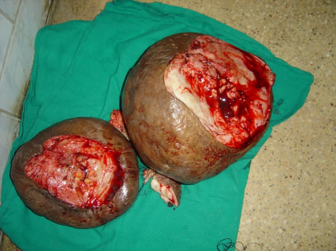
Examination of the patient revealed malnourishment and pallor. Doctors observed two huge swellings both on her right and left vulva. Furthermore, the left vulvar swelling extended below her knees while standing. The swellings made walking extremely difficult for the patient and restricted her mobility. Doctors also noticed healed scars on her cervical and inguinal regions
The doctors also took her blood samples which revealed severe anaemia with a haemoglobin level of 6 gm/dl. However, her leukocyte count came out normal. Furthermore, all of her other tests such as X-rays for chest and abdomen and pap smear etc. came out normal too.
Surgical Intervention
After going through an extensive round of tests and investigations, the doctors decided on surgery. After stabilizing her haemoglobin levels, they took her to the OR. They removed most parts of the lympho-fatty tissue to reduce the size of the swellings. However, they left out some fat tissue to retain the natural bulging appearance of the labia. During the surgery and in the immediate postoperative period, they faced leakage of lymph but managed it accordingly. Additionally, the post-op period was not very significant. The patient did develop a seroma on a follow-up presentation. However, it was treated on-site with aspiration and pressure bandaging. Over the next 6 years, doctors kept the patient on regular follow-up and she showed a swift recovery.
Vulvar Elephantiasis: A Comprehensive Insight
Elephantiasis, also known as lymphedema, refers to a swelling that develops due to the accumulation of lymph in the interstitial tissue. Our bodies have a lymphatic system to carry the fluid that leaks from the blood vessels back to circulation. The leaked fluid is called lymph and insufficiency of the lymphatic system leads to a build-up of this fluid in the interstitial spaces. This results in swelling of the tissues and oedema. Initially, oedema displays a pitting nature. Then over time, a local inflammatory reaction takes place in the affected regions followed by the deposition of fibrotic and fatty tissue elements. At this stage, oedema becomes non-pitting and the entire region acquires a gigantic appearance hence called elephantiasis.
Lymphedema can be either primary or secondary. When the cause behind the diagnosis is unknown, it is referred to as primary lymphedema. On the other hand, secondary lymphedema occurs in response to an established cause such as lymphatic insufficiency due to the removal of regional lymph nodes. Other causes include direct trauma or infestation of the lymphatics by parasites such as Wucheria bancrofti. Overall, secondary lymphedema shows a greater frequency and is easier to treat than primary lymphedema.
Vulvar Elephantiasis and TB
There have been a few case reports on vulvar TB caused by direct infiltration of the vulva by the tuberculous bacteria. These cases have also documented vulvar swellings as presenting complaints. However, vulvar elephantiasis due to a previous history of lymph node TB and destruction of inguinal lymphatics is very rare. In this patient, the vulvar tissue turned out negative for tuberculosis histology, which suggests that there was no direct invasion of the vulva by TB. However, a positive history of lymph node TB in the affected region along with evidence of healed scars suggests TB-induced lymphatic destruction as the only cause.
Lymph node TB destroys the lymphatics which significantly compromises the lymphatic drainage system. Failure to carry back lymph results in lymphedema and elephantiasis. Although rare, this case highlights the importance of paying special attention to such conditions so as to initiate timely management and avoid preventable complications.


