Case of accessory and cavitated uterine mass in 14-year-old.
This article describes the case of a 14-year-old girl who presented to the with chronic recurrent pelvic pain. The patient said that her pain aggravated every month during her menstrual cycle. Her flow, however, was normal and cycle was regular. There was no significant personal or family history, nor did she have any previous sexual history. She was diagnosed with an adnexal mass causing the pelvic pain.
She had been prescribed different kinds of anti-inflammatory medications in the past two years. The patient was also on oral contraceptive pills for 2 months. The patient did not show any significant improvement with the medications. Physical examinations showed that she was a well-developed girl with normal vital signs. She had normal pubertal height of 160 centimeters (percentile: 47%) and weight of 48 kilograms (percentile: 43%), and normal adrenarche and thelarche (stage 4).
Investigation and examination
Doctors examined the external genitalia which showed no evidence of imperforated hymen. Abdominal examination showed no detectable mass. For further examination, the patient was advised an ultrasonography. It showed a normal size and myometrial echogenicity of the uterus. Endometrial thickness was also seen to be normal. Examination showed an adnexal mass, measuring 35 x 30 mm. Similarly, the mass was left-sided heterogenous, mostly hyperechoic with close contact to the left ovary.
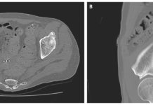
Colour doppler was evident of some internal vascularity. In addition, ultrasound showed a small right-sided ovarian cyst. Imaging of the cervix and vaginal canal showed no significant findings. To evaluate the lesion, doctors advised a pelvic MRI which showed a well-defined heterogenous mass with high signal intensity at T2-weighted images and low-to-intermediate intensity on T-1 weighted images. There was significant enhancement after administration of gadolinium. The mass was on the left side of the pelvic cavity near the uterus and left ovary. There were no signs of pelvic lymphadenopathy or ascites.
Doctors suspected paraovarian mass or myoma.
A laparotomy was also done which showed a well-defined fleshy mass on the left side of the pelvic cavity. The mass was located under the right ligament with no adhesion to the surrounding structures. In addition close to but separated from the left ovary. Both ovaries were normal. However, a right-sided ovarian cyst was notable with no endometrial implants. Examination showed that the uterus was completely separated from the mentioned mass. The mass was successfully removed with complete excision.
A chocolate-coloured fluid flowed from the mass. The patient had an uneventful recovery period and was discharged the next day. In addition, she was asymptomatic even after one-year of follow-up. Pathological evaluation of the pelvic mass led to the diagnosis of an adnexal mass.
References
Pelvic Pain and Adnexal Mass: Be Aware of Accessory and Cavitated Uterine Mass https://www.ncbi.nlm.nih.gov/pmc/articles/PMC7892247/


