Pneumothorax – delayed presentation of covid-19
This article describes the case of a 59-year-old who developed spontaneous pneumothorax after recovery from covid-19. The patient tested positive for covid-19 in January. However, did not require any hospitalisation or ventilation and recovered with supportive care. 16 days post recovery, the patient presented to the emergency with complaints of stabbing pain in the right chest and sudden shortness of breath.
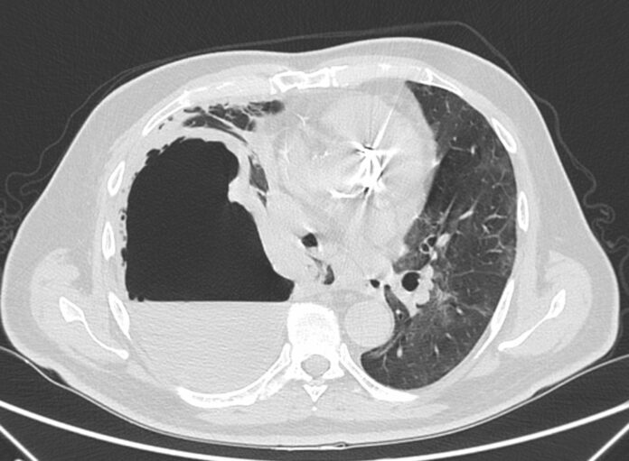
On examination, his blood pressure was 120/80 mm Hg with a pulse of 130/min. His oxygen saturation was 91% on room air. Doctors advised blood tests which were significant for elevated PCR. Whereas PCR (RT-PCR) nasopharyngeal swab was negative for SARS-CoV-2 RNA. A CT scan was also performed which showed a massive pneuothorax on the right side. In addition to this, the loves of the right lung showed extensive collapsed portions of pulmonary parenchyma. The scan was also significant for consequent mediastinum shift towards the left. Similarly, the left lung also showed ground-glass opacities.
Drainage was placed in the right chest and he was given oxygen with a venturi mask. After treatment he showed significant clinical and radiological improvement.
Chest CT findings were consistent with pulmonary involvement of SARS-CoV-2 infection
Common findings of covid-19 pneumonia include ground-glass opacities and at a later stage show multi-lobular, bilateral and peripheral distribution. The CT scan also shows consolidation, paving pattern and air bronchogram. Other rare findings include pericardial and pleural effusion. Spontaneous pneumothorax is reported in only 1% of cases with a higher predilection in men of 88%. According to some authors, “the main cause of pneumothorax in patients with COVID-19 are cystic lesions, which could occur as a result of barotrauma due to mechanical ventilation, and alveolar damage due to coughing, which causes an increase in chest pressure and ultimately an alveolar breach”.
Pneumonia because of covid-19 can cause alveolar swelling, giant bullae, fibrosis, subpleural infiltrates and inflammation in the alveolar septa. The conditions can cause parenchymal damage with pneumothorax and alveolar rupture. Literature review shows that pneumothorax is uncommon and there is a higher risk during active SARS-CoV-2 infection. However, given the case of the patient, severe complications are possible even when the infection is overcome.
References
Spontaneous pneumothorax as a delayed complication after recovery from COVID-19 https://casereports.bmj.com/content/14/5/e243578


