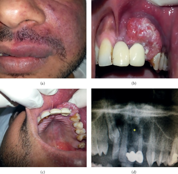
Case of peripheral ameloblastoma in upper gingiva with port wine stain in 37-year-old patient.
A 37-year-old man presented to the University Dental Hospital of Sharjah with a soft tissue mass on the left anterior part of the maxillary gingiva. The lesion has a history of 6 months. The patient’s history further revealed that the lesion had grown in size during this period with frequent episodes of bleeding in the absence of trauma. He also had a congenital port-wine stain on his left facial skin along the maxillary division of the trigeminal nerve.
Examination
Intraoral examination showed a labial painless bluish red mass measuring 3 x 4 cm in diameter. The mass was firm, pedunculated, nontender and nonpulsatile on examination. It extended from the upper right central incisor to the upper left canine. In addition, the overlying oral mucosa showed some degree of keratinisation with ulcers that bled easily. The left lateral incisor, however, was nonvital, mobile and crowned. The upper left canine was vital on the vitality test. Whereas the upper right lateral incisor was firm.
The patient’s oral hygiene was quite poor and he suffered from generalised chronic periodontitis. There was no sign of any regional lymphadenopathy. The facial port wine stain extended intraorally, involving the upper left labial mucosa, buccal gingiva and left side of the hard and soft palates.
Panoramic radiograph showed a well-demarcated periapical radiolucency in the alveolar region which extended from the root of the upper right central incisor to the upper left canine, with a non vital upper left lateral incisor. A supernumerary tooth was also detected distal to the root of the upper left central incisor with a radiolucent lesion. In addition, an impacted right maxillary canine was also present. Although, irrelevant to the lesion. The nasal cavity and maxillary sinus were normal and free from the lesion.
The patient refused to get the lesion excised under general anaesthesia and consented to only getting the soft tissue lesion removed.
References
Peripheral Ameloblastoma of Upper Gingiva in a Patient with Port-Wine Stain https://www.ncbi.nlm.nih.gov/pmc/articles/PMC7238321/


