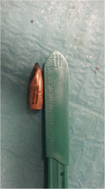
26-year-old Libyan man presented to a recovery centre in Italy with a firearm wound.
A 26-year-old Libyan man presented to a recovery centre in Italy with a firearm wound in his abdominal wall from the left to ride side with no exit wound. He was running, seeking refuge when the bullet hit him. The bullet had an entrance orifice of 3 cm diameter at the left lumbar paravertebral region. The victim was a clandestine refuge from Libya who spent 20 days crossing the Mediterranean sea.
He was admitted to a a recovery centre and then to a local hospital in Italy. He presented to the hospital in a good general condition and did not show any signs of hemodynamic shock.
An abdominal radiograph was performed which showed a bullet located anterior to the second lumbar vertebra. The tip of the bullet rotated in the upright direction associated with fractures of the second and third lumbar vertebrae.

The patient was immediately transferred to the neurosurgery department of the hospital. The patient was hemodynamically stable when brought to the neurosurgery department. Preoperative CT angiography showed that the bullet had partially penetrated into the aortic wall at the level of the left renal artery. There were no signs of surrounding hematoma or active blood loss.

The patient was admitted with a Glasgow Coma Scale of 15, hyperthesia, hypothenia and lumbar pain. The patient was prepared for surgical repair and the aorta was accessed through a lobotomy retroperitoneally. The. bullet was visualised at the level of the left renal artery, partially penetrating the left latero-posterior aortic wall. Furthermore, the bullet crossed the lumbar spine, fracturing the second and third lumbar vertebrae, penetrating half of the aortic wall. There was no visceral injury or surrounding hematoma.
The patient’s postoperative period was uneventful. He was discharged after 6 days of the surgical procedure with no neurological symptoms.
References
Penetrating aortic injury left untreated for 20 days: a case report https://www.ncbi.nlm.nih.gov/pmc/articles/PMC5787315/


