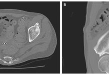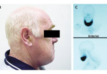
67-year-old female patient was diagnosed with metastatic adenocarcinoma 23 years after breast cancer diagnosis.
67 year old female patient with a history of breast cancer presented with complete loss of vision in her left eye with a history of 3 months. This was preceded by a 3-month history of progressive blurring of vision. The patient was diagnosed with base of skull metastatic adenocarcinoma 23 years after her breast cancer diagnosis.
History and examination
The patient was diagnosed with left vocal cord palsy a year before. She presented with the symptoms of hoarseness. In addition, her medical history revealed breast cancer for which she underwent left mastectomy and axillary clearance. She was on radiotherapy till 1992.
Clinical examination of the patient showed loss of vision and absent corneal reflex, rectus palsy in the right eye, left vocal cord palsy and hypoglossal nerve palsy. Endoscopy showed a well-encapsulated mass in the nasal cavity’s superior aspect. There were no contralateral breast masses palpable nor any axillary lymphadenopathy. The findings showed no evidence of malignancy. Radiological and blood investigations were also normal. A contrast-enhanced computed tomography of the base of the skull, paranasal sinuses and neck region was performed. The scan was remarkable of a large, lobulated, heterogeneously enhancing mass at the sphenoid bone, measuring 5.7 × 6.4 × 4.8 cm. Another similar lesion was also seen within the left jugular fossa, measuring 2.8 × 1.9 × 2.5 cm.
Gadolinium-based contrast magnetic resonance imaging (MRI) also showed an anterior skull base tumour that surrounded the olfactory nerve and eroded the sphenoid sinus. A similar lesion was seen within the left jugular foramen. Histopathological analysis confirmed metastatic adenocarcinoma. A bone scan after the biopsy was also performed a month after the biopsy, showing local bony destruction that involved the clivus, bilateral petrous, bilateral anterior arc, and lateral mass of C1 vertebra.
Treatment
The initial treatment plan included radiotherapy and chemotherapy. The patient completed 10 cycles of radiotherapy (total of 30Gy) and completed 6 cycles of chemotherapy. She was later prescribed an aromatase inhibitor and calcium supplement. However, 20 months after the initial investigation, repeat CT of the base of the skull did not show any significant changes in the size and extension of the previously seen mass. The mass in the left jugular fossa was not visible. In addition, the left vocal cord deviated medially because of recurrent laryngeal nerve palsy secondary to vagus nerve compromise.
The patient complained of dysphagia 10 months after treatment. Therefore, she was referred for percutaneous endoscopic gastrostomy (PEG) tube insertion. 15 months after completion of chemotherapy and radiation, the patient was generally well with no history of epistaxis. Although, she was still on PEG tube feeding. Moreover, she was solely using her left eye because of not regaining vision in her left eye.
References
Base of Skull Metastatic Adenocarcinoma from the Breast 23 Years after the Primary Diagnosis https://www.ncbi.nlm.nih.gov/pmc/articles/PMC7414367/


