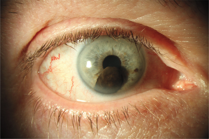
Case of 57-year-old patient with 4-month history of pain and decreased vision in one eye.
This article describes the case of a 57-year-old male patient with iridociliary melanoma with secondary glaucoma. The patient presented to the ophthalmology clinic with a 4-month history of chronic pain associated with decreased vision in the right eye. According to the patient, when he used his affected eye, he could only see at a distance of 1 m. Similarly, he was only able to count his fingers with the affected eye.
Examination was remarkable of a pigmented nodule located in the inferior part of the iris in addition to smaller pigmented lesions. His intraocular pressure was 63 mm Hg (reference range, 10 to 21). Whereas fundus examination showed cupping of the right optic disk. Similarly, as seen in the ocular ultrasonography, the mass involved the iris and ciliary body. The findings were consistent with the diagnosis of iridociliary melanoma with secondary glaucoma.
Iridociliary melanoma
Iris and ciliary body melanoma is defined as an aggressive tumour that only presents symptoms in its advanced stages. Doctors often discover the melanoma incidentally during routine examination. Treatment includes monitoring the tumour, radiotherapy and enucleation.
Doctors performed a primary enucleation of the right globe. Histopathology of the tumour confirmed the diagnosis. Evaluation for extraocular metastases was negative. Early detection of ocular melanomas allows less invasive treatment, surgeries or radiation therapy.
The patient was advised regular follow-up. However, he did not follow-up after surgery.
References
Iridociliary Melanoma https://www.nejm.org/doi/full/10.1056/NEJMicm2012968


