Woman develops pyogenic granuloma on her left thigh!
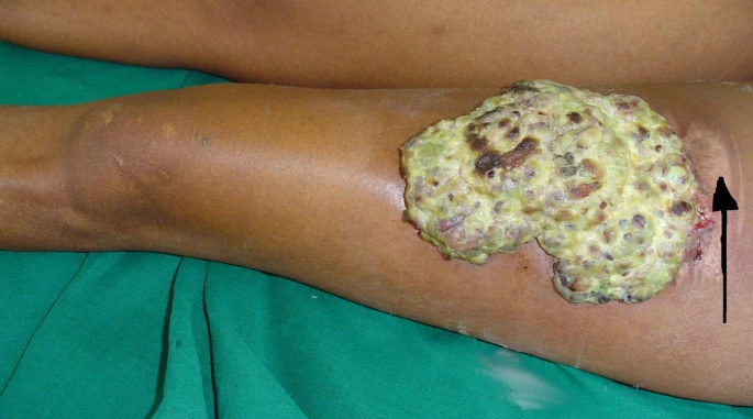
A 28 year old woman presented to the hospital with an ulcerated mass on her left thigh. She went through an accident during which she avulsed the anteriomedial part of her left leg, 9 months prior to her presentation. The initial wound was managed for infection through regular dressings and medication. However after 3 months, doctors proceeded with debridement and replacement of the defect with a split skin thickness graft. They used an autograft from the patient’s own left thigh. Although, the original defect showed good healing after graft placement, the donor site did not heal. It developed ulceration with purulent discharge coming out of the wound.
The ulcerated mass on the patient’s thigh was very painful. She had remained bedridden for two months because of the pain. Moreover, she complained of foul smelling discharge and bleeding from the wound as well.
Examination of the Ulcerated Mass
On examination, the patient’s wound was covered by a creamy tissue. Doctors noticed blood clots on the edges of the wound indicative of previous episodes of bleeding. In addition, the proximal margins became continuous with the adjacent skin. Overall, the doctors did not notice any adjacent tissue abnormalities or discolourations in the adjacent skin.
As a treatment, doctors excised the mass with appropriate margins and used another split skin graft to cover the tissue defect. They sent the excised specimen for biopsy. The results revealed pyogenic granuloma. Results also indicated the presence of bacteria on the surface of the wound only. It was clear of any fungal infiltrates.
What is Pyogenic Granuloma?
Pyogenic granuloma, called a lobular capillary hemangioma, refers to a benign condition that affects the skin and mucosal surfaces. It develops as small red lobules, bumps or growths that bleed very easily because they contain a lot of blood vessels. Although its usual site of occurrence is skin and mucosa of the proximal GIT, it can also develop intravenously and in the distal gastrointestinal tract such as in the colon and rectum. However, regardless of where it develops, it always remains benign and offers a good prognosis.
Causes of Pyogenic Granuloma
Pyogenic granuloma develops more frequently in children and young adults but that does not mean that it can not develop in other age groups. It can even develop in pregnant females where it likely sets in due to hormonal changes. Anyhow, there are multiple causes for capillary hemangioma. It can develop at the site of an injury or can develop as a result of trauma such as excessive scratching after bug bites. People on certain medications such as birth control pills can also develop a pyogenic granuloma. Moreover, people who are immunologically compromised are also at high risk for developing a capillary hemangioma.
Stages of Development
During the initial course of its development, this hemangioma develops aggressively and increases in size for a few weeks. The growth then slows down and it resorts to a small reddish nodule not greater than two centimetres. At first, the nodule is smooth and shiny whereas, with time, it becomes rough and starts bleeding even with mild traumas.
Doctors identify two other conditions that mimic PGs in both their clinical presentation and histological characteristics. They are bacillary angiomatosis and verruga peruana. The former involves widespread vascular lesions whereas the later develops as crops of vascular nodules in immunologically incompetent people. Both of these conditions set in after a Bartonella infection and since they resemble PG so much, doctors often hypothesize that the same infection can be the cause of PG as well. However, we do not have any conclusive evidence to support this hypothesis.
Pyogenic Granuloma and HIV
Pyogenic granulomas can develop at the graft site in patients who are HIV positive and receive a skin graft, as in our patients. However, they usually develop in the form of multiple lesions and are less than 2 cm in size. We have never seen a solitary presentation of pyogenic granuloma with a size as big as something almost covering up the entire thigh. A rare case by all means!


