Case study: cystic echinococcosis
This article describes the case of a giant isolated splenic hydatid cyst, secondary to cystic echinococcosis, in a 49-year-old Balkan woman. The patient presented with a 6-month history of left upper quadrant pain without tenderness. Her past medical history was insignificant. Physical examination showed a mass occupying the left upper abdomen. Doctors further advised an ultrasound that showed an uncertain cystic mass with solid components. CT scan of the abdomen and pelvis showed a septated splenic mass, measuring 22 x 18 x 13 cm.
The findings further showed that the voluminous cyst was displacing the right kidney, stomach and pancreas. There were no signs of liver cysts and inflammatory markers were normal. On suspicion of cystic echinococcosis, the patient was referred to the clinic. However, the patient’s medical history revealed that she has no contact with animals in her hometown. Doctors further advised a CT scan of the lungs and MRI of the brain, which did not show any additional lesions. The echinococcus IgG enzyme-linked immunosorbent assay (ELISA) test was also negative.
Treatment and prognosis
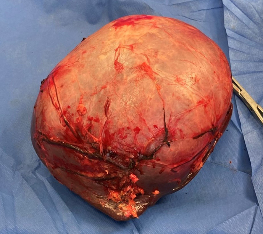
Treatment included a splenectomy because of the size of the cyst and compression of the splenic parenchyma.
2 weeks before the patient was admitted she was vaccinated against pneumococcus, meningococcus and influenza. Doctors further referred the patient for an anti-infective albendazole (ABZ) treatment. Her postoperative period was uneventful and she was discharged on the 8th day after the procedure. In addition, she was on adjuvant treatment with ABZ for a month.
Histopathological analysis of the lesion showed a parasitic cyst with an outer pericyst, weighing 3.05 kg. Cyst fluid cytology was consistent with the diagnosis of cystic echinococcosis. The patient was called for a follow-up after 6 months of the procedure. Abdominal ultrasound, echocardiogram and a chest X-ray was done which showed no signs of recurrence of infection.
A hydatid cyst is a common presentation of echinococcosis in humans. The disease is most commonly localised in the liver and lungs, however, other organs can also be affected because of cystic dissemination. Isolated involvement of the spleen is rare, as with this case report.
Source: American Journal of Case Reports


