Helmholtz Munich, the Technical University of Munich (TUM), and the Medical University of Vienna have developed an innovative imaging technique known as “O2E”. That enables clinics to detect esophageal cancer with unparalleled precision.
The study, published in Nature Biomedical Engineering, shows that this novel endoscopic technique detects even the smallest diseased tissue changes, resulting in much improved early detection and diagnosis.
Esophageal cancer is one of the worst tumors; when identified at an advanced stage. The survival rate is barely around 10%. However, when found early, around 90% of patients survive. The new O2E technique could help detect alterations in esophageal tissue at an earlier stage.
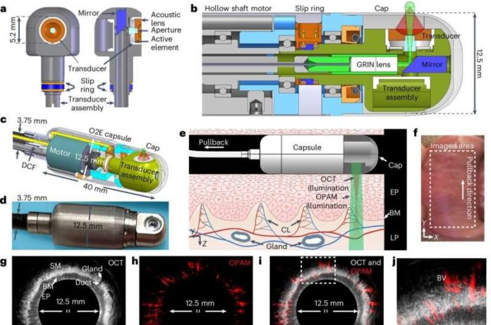
O2E is a new type of endoscopic technology that combines two imaging techniques. While optical coherence tomography is particularly excellent at catching tissue features, optoacoustic imaging, which stimulates tissue with light pulses and detects ultrasonic signals as a result of illumination, can see even the smallest blood veins in deeper tissue layers.
By combining these methods, high-resolution 3D pictures of tissue structure and function in the esophagus are produced. Both sensors are combined into an endoscopic capsule, which scans the tissue from every angle.
Prof. Vasilis Ntziachristos, Director at the Institute of Biological and Medical Imaging at Helmholtz Munich and Chair at TUM said
“Our dual imaging system uncovers critical features of early cancer lesions, including microscopic structural changes beneath the mucosal surface and subtle microvascular alterations within the cancerous tissue, that previous methods were unable to detect,”
In their pilot investigation, the researchers analyzed animal esophaguses as well as tissue samples from individuals with Barrett’s esophagus, a precursor to esophageal cancer. They successfully distinguished healthy tissue from tissue with aberrant cellular alterations, precancerous stages, and malignant tumors.
Initial proof-of-principle testing were conducted on a volunteer’s inner lip, which shares tissue features with the esophagus.
Dr. Qian Li, first author of the study from the Medical University Vienna said
We also plan to integrate confocal endomicroscopy—a technique that provides high-resolution, real-time visualization of cellular structures—to enable more detailed analysis during examinations,”


