Total Resorption of a Chronic L4-L5 Disc Extrusion After Application of the Atlasprofilax Method
Invertebral disc extrusion occurs in up to 75% patients with symptomatic extruded lumbar discs. It has been described in literature by various authors. The Atlasprofilax method has been shown to favour osteo-musculo-articular rehabilitation process. It mainly impacts on 2 levels, including the bone anatomy of the atlano-occipital joint and fasciae located in the cervical region. This article describes the case of a 42-year-old male patient diagnosed with dehydration of L2-L3 to L5-S1 via MRI. The patient’s disc was seen bulging at L2-L3 and L3-L4 with disc extrusion at L4-L5.
Doctors suggested that altering the cervical musculature and deep fascia could lead to the development of lumbar disc abnormalities. The paper further proposed that the improvements in symptoms and imaging findings in this patient are because of a reduction in the asymmetric distribution of forces.
Case Report: Atlasprofilax Method
A 42-year-old male patient was diagnosed with L2-L2 to L5-S1 intervertebral discs and bulging at L2-L3 and L3-L4 and disc extrusion at L4-L5. The diagnosis was confirmed through an MRI. Doctors advised a one-time neuromuscular treatment called the Atlasprofliax method, to the suboccipital region. 6 months later, doctors referred the patient for another MRI and the L4-L5 disc extrusion had resorbed completely. Prior to Atlasprofilax, the patient suffered from bilateral trapezius pain, lower limb dysesthesias with pain and numbness in calves, gait claudication, right sciatica, constant chronic low back pain and right brachialgia. A week later the sciatica symptoms disappeared and did not recur.
The patient was called back for a follow-up 5 years later and the remaining symptoms had also shown further improvement. In conclusion, it was seen that the reduction of the asymmetric distribution of forces and elastic load shedding as a result of the Atlasprofilax treatment showed improvement. In both the symptoms and imaging findings of the patient.
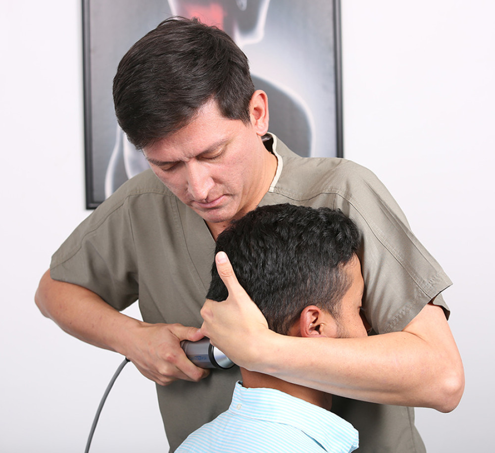
Intervention
The patient was referred for the intervention via Atlasprofilax method in a single session. In this method, the doctors work with myofascial structures of the neck using a device. The device uses the principles of mechano-transduction by vibropres-sure at special frequencies. In addition, the frequencies are applied to the several keypoints of the suboccipital area to stimulate certain muscle and facial receptors. The aim of the procedure is to allow a deep mechano-transduction effect on the suboccipital muscles.
The study objectives included observing imaging improvement reduction of the size and morphology of the herniated disc by means of an MRI. In addition to associated clinical manifestations. The size and morphology of the affected extruded discs were analyzed at the imaging level and taken as endpoints. This was to check if there was any improvement, such as disc resorption.
The result of the pain at the clinical level was used to determine the improvement in life quality and therapy outcome satisfaction using the PGIC scale.
Radiological Findings
The radiological findings were as follows:
“There is bulging of the annulus fibrosus of the intervertebral discs of L3–L4 and L4–L5 with presence of tearing of the annulus fibrosus in the posterior aspect of the intervertebral disc of L4–L5, this bulging associated with fascicular degenerative changes moderately decreases the width of the spinal canal in these locations, without relevant associated narrow, central, or lateral canal.”
Furthermore, “The right paracentral disc herniation visualized in the previous study is not observed and the width of the spinal canal has improved for this reason in this location. Currently, the right paracentral disc herniation L4–L5 visualized in the previous study is not observed. In the present study there is bulging of the fibrous ring L3–L4 and L4–L5 with moderate decrease in the amplitude of the spinal canal, which, however, has improved with respect to the previous study.”
In conclusion, a clear improvement was seen in the quality of life according to the PGIC scale.
Source: American Journal of Case Reports


