Pneumococcal empyema
This article describes the case of a 4-year-old girl who was brough to the emergency with fever, cough and lethargy with a history of 4 days. Her medical history revealed that she was up to date with all her vaccinations. كازينو اونلاين Three weeks earlier doctors presumed that she had a respiratory tract infection with SARS-CoV-2, however, tested negative at the time. Physical examination showed a heart rate of 136 beats per minute, oxygen saturation of 92% and respiratory rate of 32 breaths per minute. موقع مراهنات رياضية Doctors diagnosed her with pneumococcal empyema.
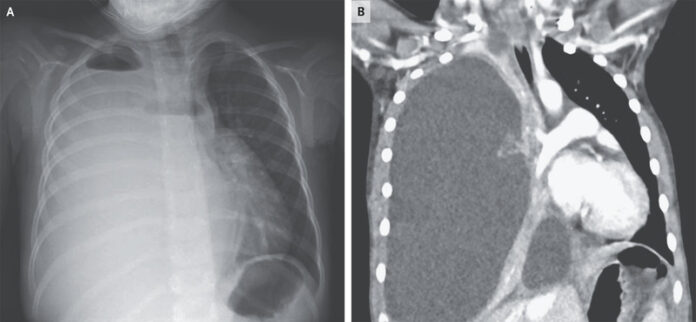
Auscultation of the lungs showed absent breath sounds on the right side, whereas the left side showed diminished breath sounds. Doctor advised further laboratory tests which showed a leukocyte count of 25,300 per cubic millimeter (reference range, 5000 to 15,500) and 88% neutrophils. Chest radiography further showed opacification of the right hemithorax, with apical fluid levels consistent with the presence of hydropneumothorax. بلاك جاك اون لاين
Diagnosis and treatment
A computed tomography of the chest was also performed which showed right lung collapse because of massive effusion and mediastinal shift towards the left. The left lung parenchyma, however, appeared to be normal with no abscesses or cysts. A chest tube was placed, and 1 litre of cloudy fluid was removed. The pleural fluid cultures were positive for streptococcus pneumonia.
Community acquired pneumonia is one of the most common infections in children. Similarly, there are 24 to 40 cases of pneumonia reported per 1,000 children in Europe and North America, each year. When diagnosing pneumonia, patient’s history, physical examination, laboratory tests and chest radiographs play an important role. Even in fully vaccinated children, pneumococcus is an important cause of postviral pneumonia and empyema. The patient was treated with ceftriaxone for 14 days. At 3-months follow-up, the patient showed full recovery and repeat chest radiograph was normal.
References
Pneumococcal Empyema https://www.nejm.org/doi/full/10.1056/NEJMicm2035551


