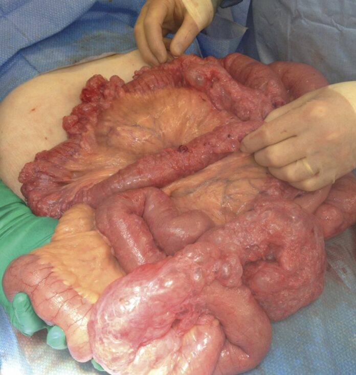
Pneumatosis intestinalis in 88-year-old with complaints of abdominal pain and vomiting
This article describes the case of an 88-year-old who presented with complaints of severe abdominal pain and vomiting. Doctors advised a chest x-ray which showed free intraperitoneal air. The findings were also consistent with small bowel obstruction. For further investigation a CT scan was advised which showed free intraperitoneal gas. Based on these findings, doctors diagnosed the patient with pneumatosis intestinalis.
Pneumatosis is the presence of gas in the bowel wall, which is often identified through abdominal radiographs or computed tomography (CT) scans. The cause of the condition range from benign to fulminant. It is often seen to be associated with ischemia, especially in case of portomesentlric venous gas. It is also often seen in other conditions, for example, AIDS, amyloidosis, leukemia, celiac disease, infectious enteritis, connective tissue disorders and chronic obstructive pulmonary disease. The diagnosis of PI in neonates is associated with necrotising enterocolitis and has a high mortality rate.
The condition was initially described by Du Vernoi in 1783
Since then, it has been referred to with multiple pseudonyms. In addition, the incidence of the condition has increased since the use of CT for intraabdominal pathology. It is also believed that the incidence may have increased because of iatrogenic causes, including the instrumentation of the gastrointestinal tract and prescribed medications.
Surgical intervention may be required in patients with acute abdomen, ischaemic bowel and evidence of obstruction. PI is commonly identified on routine radiographs of the abdomen. During endoscopy, submucosal cysts may also be identified occasionally. The cysts may appear similar to polyps. The polyps can be examined at biopsy for identifying signs of inflammation. The gas is usually seen collecting peripherally in the lumen of the bowel and around contrast or fecal material. This gas can trigger pneumatosis which is visible on CT scans.
Source: BMJ


