Incidental finding of inferior vena cava
This article describes the case of a patient with blunt abdominal trauma, which led to an incidental finding of a duplicate inferior vena cava. A 36-year-old man presented to the emergency after he got into a motor vehicle collision. On the patient’s arrival at the emergency, the ATLS protocol was followed. In addition, the patient was stable with a GCS score of 15.
The patient complained of the right flank and hip pain. The examination was consistent with the ‘seatbelt sign’, a clinical and radiological sign characterised by the presence of ecchymosis or abraded skin extending along the abdomen after a motor vehicle accident.
Investigations and findings
Doctors further advised focused assessment with sonography for trauma (FAST), which was negative. A CT was also performed subsequently which showed the presence of bilateral pre-nephritic fat in the abdomen with mesenteric fat. In addition, a small amount of free fluid was also evident in the right iliac fossa. The findings were consistent with a possible mesenteric injury. The findings were also significant for a duplicate inferior vena cava which was unrelated to the patient’s presentation.
Doctors admitted the patient for observation and conservative management. Similarly, further advised serial abdominal examinations. However, the patient was hemodynamically stable despite his complaint of lower-quadrant pain. He was put on a liquid diet. On the 5th day, he complained of increased severity of pain and was referred for a repeated CT. The CT was significant for interval thickening of the ileal loops wall. Moreover, incidentally revealed a duplicate inferior vena cava.
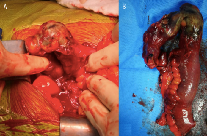
Other findings included a lack of wall enhancement in the ileum with sub-segmental ischemia. The distal ileal branch was poorly pacified and irregular. In addition, there was an evident increase in the amount of free fluid, with no evidence of free air.
Doctors referred the patient for an urgent laparotomy. The patient had an uneventful recovery and was discharged on the 13th postoperative day. The patient returned for a follow-up to the outpatient clinic.
Source: American Journal of Case Reports


