Cancer is a category of chronic diseases that cause abnormal growths in the body called tumors. They can be classified as either benign (where cells don’t spread but the mass grows big enough to cause distress to organs) or malignant (where the cells spread to other parts of the body). These tumors can cause serious health problems by exhausting oxygen and nutrients so that normal body cells can’t receive them.
While research has made significant advances in treating cancer, there is still no perfect cure for it. Usually, the plan of action includes surgically excising the cancer, followed by radio and chemotherapy. However, malignant cancers do not have clear boundaries that separate them from normal tissues. This makes it hard to judge how much tissue to remove. This is a serious disadvantage because if the surgeon fails to remove all the malignant tissue, the cancer can recur.
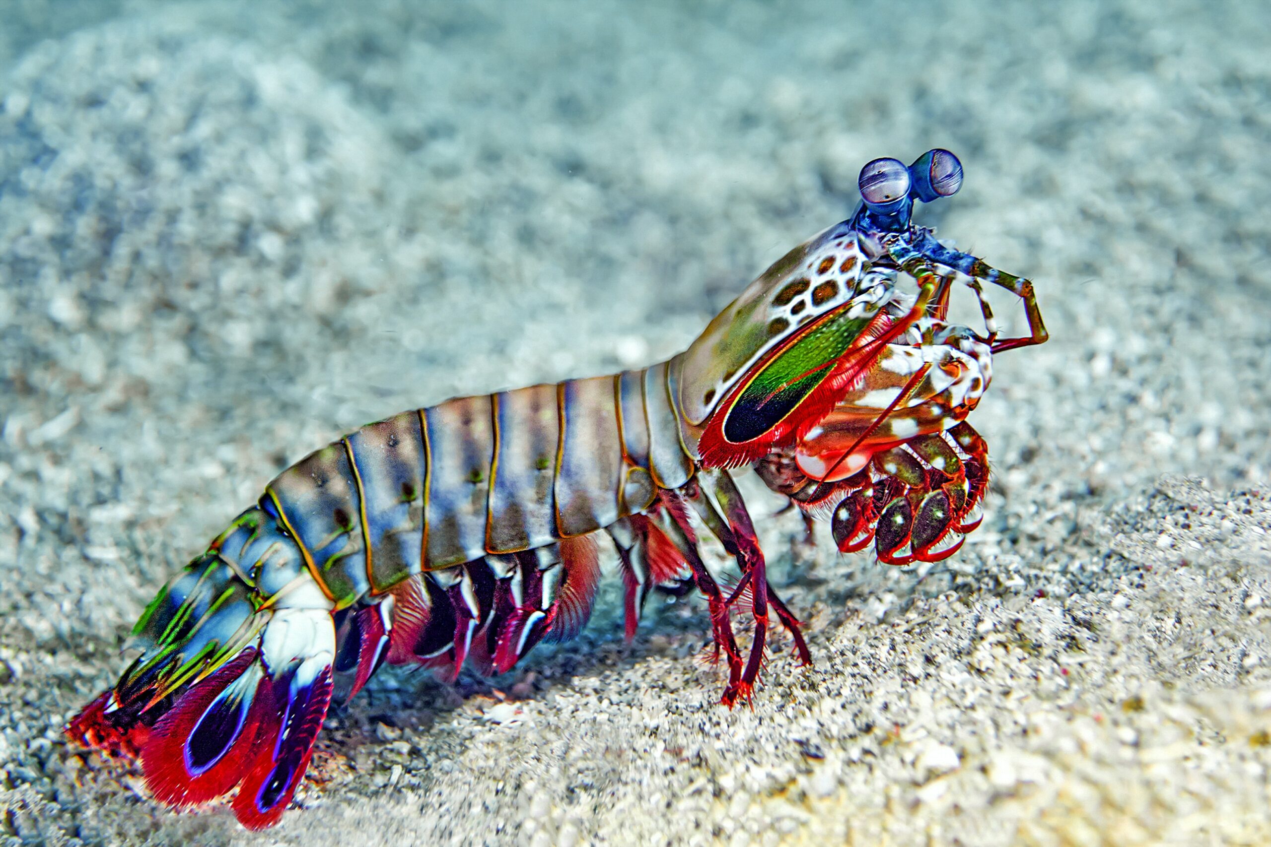
Recently, a team from North Western University have designed a new camera to help surgeons distinguish cancerous and healthy cells. They took inspiration from a unique sea creature: the mantis shrimp.
A New way to look at Cancer
According to lead author Steven Blair from the University of Illinois Urbana-Champaign “The mantis shrimp has these incredible eyes. Humans perceive three colors – red, green, and blue – because of a single layer of light-sensitive cone cells that line our retina, but the mantis shrimp perceives upward of 12 colors thanks to the stacks of light-sensitive cells at the tip of its eye. The mantis shrimp can thus see things that humans can’t imagine – and it does so in a fraction of the space.”
In the study published in Science Translational Medicine, the researchers describe a camera that sees six colors. The visible light spectrum makes up 3 of these colors, while the other 3 can detect near-infrared fluorescence. To use this technique, doctors will inject the patients with a chemical. This causes the target tumor to shine in near-infrared light, making the cancerous tissue easy to distinguish.
According to the study, the researchers have already tested this camera on mice that bore prostate cancer cells. They were able to accurately distinguish between healthy and cancerous cells in 92% of the cases. The camera also improved tumor resection in head and neck cancer patients. It also helped surgeons accurately map out and remove affected lymph nodes in 18 breast cancer patients.
Goran Kondov, a professor and chief surgeon from North Macedonia demonstrated this technology in the operating room. He said, “The combination of this bioinspired camera and emerging tumor-targeted drugs will ensure that surgeons leave no cancer cells behind in the patient’s body. This additional set of eyes will help prevent recurrence of the disease, providing patients a quicker and easier path to recovery. And the device can potentially be manufactured at low cost since it is so simple, making it accessible to hospitals around the world.”


