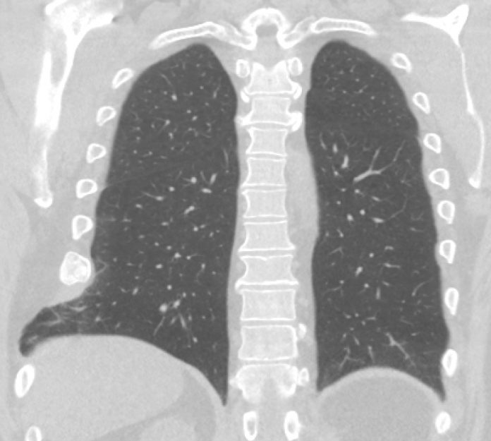
A 54-year-old male presented to the clinic with complaints of a palpable mass on the right side of his chest associated with pain and discomfort. The patient had a history of heavy smoking. He denied any recent trauma or any event that he could relate to the appearance of mass.
Although he mentioned an accident that happened around 4 years ago, the patient had fallen from a considerable height around 4 years back for which he never sought professional help as he did not suffer fatal injuries, nor had any considerable symptoms. Few years after the fall, he noticed a cracking sound from right thorax after multiple episodes of sneezing. Again, since the patient didn’t consider it to be significant, he did not seek medical attention.
Four years later, the patient presented with this palpable mass and pain in the right thoracic region, which became noticeable after considerable weight loss.
On examination, there was a noticeable, palpable bulge with a positive Valsalva maneuver. On plain radiography, the right side of the chest revealed rib fractures with callus formation of the 7th, 8th, and 9th ribs. There were no evident pulmonary abnormalities.
A computed tomography scan revealed consolidated fractures of ribs from the 7th to 9th rib on the right side with lung tissue herniating from between ribs 7 and 8 (in the image above).
It was planned to manage the patient surgically. Intraoperatively, multiple consolidated rib fractures were seen with secondary malalignment of the ribs. An old tear was identified between the intercostal musculature of the 8th and the 9th ribs through which the lung tissue was herniating. The defect in the intercostal muscle was repaired with a polypropylene mesh, and the 8th and 9th were reconstructed with a rib plate.
The surgery and the initial postoperative period were uneventful. Three weeks after the surgery, the patient complained of swelling at the surgical site. An ultrasound was performed, which revealed a large fluid collection, which was drained out. The collection was found out to be a seroma.
After the drainage, there were no further complications or signs of infection, and at the 1-year follow-up, the patient seemed to have recovered substantially.
References:
Knoef RJH, Wemeijer TM, Steenvoorde P, Groot R (2020) Spontaneous Lung Herniation after Coughing: A Case Series. Trauma Cases Rev 6:083. doi.org/10.23937/2469-5777/1510083


