Presentation: History and Examination
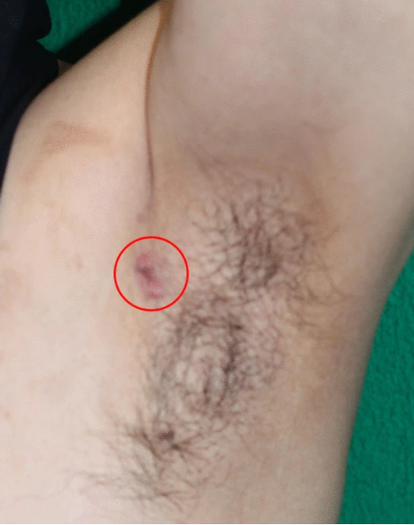
A 64-year-old man from Japan presented with a small nodule in his left axilla for 2 years. According to the patient, he noticed that the nodule gradually increased in size, which is why he visited the dermatologist. However, his medical history did not give any significant medical or family history of any such disease. Moreover, he did not give any history of smoking or drinking alcohol. The doctors performed an incisional biopsy of the axillary nodule, which came out positive for a neuroendocrine tumour. He was then referred to the oncology ward due to the biopsy findings.
On examination, a hard axillary nodule, measuring 15 mm x 10 mm was found in the left axilla. Although, no other abnormal examination findings were noted. Neurological examination was normal. The baseline blood investigations were normal, and the patient was vitally stable.
Investigations: Neuroendocrine Tumour
The doctors performed an ultrasound of the nodule and revealed a 15mm mass that was irregular with enlarged lymph nodes with normal-sized mammary glands. A CT scan was also performed which showed an irregular mass in the left axilla with enlarged lymph nodes and no evidence of metastasis. Another radiological investigation called fluorodeoxyglucose (FDG)-positron emission tomography (PET)/CT showed similar findings with no evidence of any other mass.
Management
The doctors managed the axillary tumour with wide radical excision of the axillary tumour and the axillary lymph node dissection.
The doctors administered four courses of Taxotere and cyclophosphamide (TC) chemotherapy (75 mg/m2 docetaxel and 600 mg/m2 cyclophosphamide) every 3 weeks. Moreover, endocrine therapy (20 mg/day tamoxifen) and radiotherapy of the left axilla and lymph nodes (50 Gy/25 Fr) were given after chemotherapy.
Histological findings of the Axillary Tumor
The specimen obtained from the excision of the tumour was preserved in formalin. The histological evidence of the axillary tumour revealed a yellow-white nodule present on the skin and the subcutaneous fat tissue measuring 15 x14 x 11 mm. The Hematoxylin and eosin (H&E) staining revealed a solid tumour in the fat tissue which was invading the skin.
The specimen showed dense cellular solid nests of cells with cores that consist of fibrovascular tissue. These cells were made of ovoid nuclei with salt and pepper chromatin and eosinophilic cytoplasm. The Ki-67 positive cells were present with a proliferation of the cells at a rate of 20.8%. The tumour was positive for CAM 5.2, which indicated the epithelial nature of the tumour with neuroendocrine differentiation (90% of the tumour). The tumour was ER (estrogen receptor) and PR (progesterone receptor) positive. This showed the mammary gland origin of the tumour which was confirmed by the presence of GATA3 staining. It was negative for HER2 (human epidermal growth receptor 2). Additionally, three axillary lymph nodes were metastatic, thus the stage of the tumour was labelled Stage IIA and pT1N1M0.
Prognosis
After one year since the management of the tumour, the patient is in remission and no recurrence has been noted.
Discussion: Neuroendocrine Tumour
This case of male accessory breast cancer originating from the axilla is very rare.
Male breast cancer comprises 1% of all breast cancers in the general population. It more commonly arises as an accessory breast cancer which presents as an axillary tumour. It is extremely important to prove the presence of the tumour with histological evidence. Moreover, metastatic disease from any other tumour in the body should be ruled out.
These neoplasms can be confused with endocrine mucin-producing sweat gland carcinoma (EMPSGC), however, this differential can be ruled out as these tumours are usually present in the head, neck and eyelids of older women. Moreover, radiological investigations such as CT, US and FDG-PET/CT can confirm the nature of the neoplasm.
Neuroendocrine tumours arise from neuroendocrine cells and can be found anywhere in the body. However, the most common sites for such tumours are the gut, lungs, and bronchi.
A Breast Neuroendocrine tumour (BNET) is a mixed neuroendocrine and non-neuroendocrine breast neoplasm with a non-neuroendocrine component of less than 10%. It has increased expression of ER and PR than other breast cancers and is usually categorized as a luminal A or luminal B HRE2-negative breast cancer. These tumours have IHC markers such as CK7, ER, PR, and GATA3.
Unfortunately, there are no specific management guidelines for Breast Neuroendocrine Tumors, therefore these tumours are managed with breast cancer treatment. Management options include endocrine therapy, chemotherapy and excision of the tumour. Moreover, postoperative chemotherapy, radiotherapy and endocrine therapy can be used.


