Case Report
Acanthosis nigricans (AN) is characterized by bilateral, darkly pigmented, coarse patches and plaques that appear in areas of skin folding, such as the groin, axillae, and neck. It may also affect the elbows, knees, dorsum of the hands and digits, external genital areas. And, in certain cases, the cheeks and eyelids. The first signs are a shift in skin color to brown in those with lighter skin tones. And gray in those with darker complexions, followed by an increase in dryness and coarseness. As the illness worsens, the affected skin thickens and develops a velvety texture, which is frequently accompanied by tiny, wart-like lumps.
The global incidence rate of Acanthosis nigricans varies greatly, with reports estimating a prevalence ranging from 7 to 74%, depending on criteria such as age, ethnic origin, type of AN, and the severity of the underlying pathology.
Acanthosis nigricans affects people of all ages, from birth to adulthood, with no apparent gender differences. It is linked to a variety of benign and malignant disorders, all of which follow a same pathway of enhanced keratinocyte and fibroblast proliferation. Obesity, insulin resistance-related diseases, endocrinopathies, familial AN, and specific drugs, such as corticosteroids, have all been recognized as leading causes of AN.
As the prevalence of obesity and insulin resistance rises, so will the number of instances of AN. Early identification of this skin problem is critical in preventing the advancement of underlying illnesses.
This case report describes an unusual symptom of AN, which contributes to a better knowledge of its clinical range.
Case Presentation
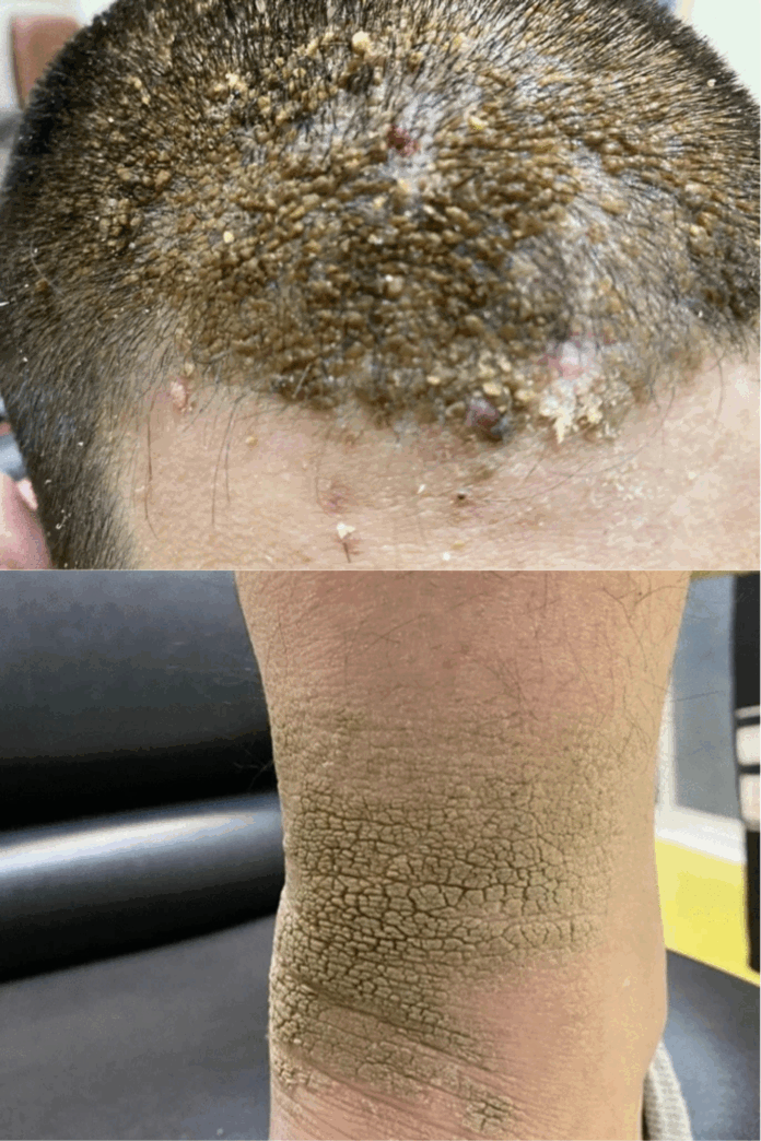
A 16-year-old Iranian male visited the dermatology clinic at Alzahra Hospital in Isfahan, Iran. He complained of lesions on his anterior scalp. Since adolescence, he had suffered from seborrheic dermatitis on his frontal scalp. Over the last four years, as the patient gained weight and developed obesity, the scalp lesions became more visible, taking on a papillomatous appearance. The patient had no history of systemic disease or pharmaceutical usage, and no similar cases were found in his family.
Physical examination revealed yellowish-brown, scaly papillomatous plaques in the frontal scalp and velvety brown plaques in the flexural areas.
Investigations:
Histopathologic examination of the scalp lesions revealed orthokeratotic hyperkeratosis, papillomatosis, and moderate acanthosis. Skin irritation and spongiosis were nonexistent. No significant parakeratosis or neutrophilic buildup were found, ruling out psoriasis. The absence of spongiosis excluded seborrheic dermatitis, and the absence of koilocytes and parakeratosis excluded warts.
An comprehensive screening for metabolic and systemic disorders often associated with AN revealed no notable findings. The histopathologic results and the presence of AN in other parts of the body, including the neck and axillae, led to the diagnosis of AN.
Treatment
Topical treatment with 6% salicylic acid shampoo, a typical keratolytic agent, was started. And administered to the scalp for three months. At the two-month follow-up appointment, the patient’s scalp lesions had improved significantly. However, the patient failed to show up for the next scheduled appointment. During a follow-up phone call, the patient reported a return of the lesions. That was attributed to inadequate long-term therapy compliance.
Discussion and Conclusion
Acanthosis nigricans (AN) is a dermatological disorder that affects the skin’s cosmetic appearance, with darkening, thicker patches caused mostly by hyperkeratosis rather than increased melanin synthesis. This hyperkeratotic state might confound the distinction of AN. Especially when it appears in unusual places, resulting in misdiagnosis and delayed detection of underlying causes such as insulin resistance. Endocrinopathies (such as obesity, insulin resistance, type 2 diabetes, and polycystic ovarian syndrome) and cancer are major comorbidities of AN. Notably, 17.6% of malignant AN instances occur before the diagnosis of malignancy. Thus, early diagnosis and long-term follow-up are critical for successful management.
The unusual incidence of AN on the scalp, particularly in areas previously afflicted by seborrheic dermatitis, raises the possibility of a link between these two illnesses, which has not been widely investigated in the literature. Sirka et al. have highlighted a probable association between AN and preceding atopic dermatitis, implying a larger relationship between diverse forms of dermatitis and AN.
Despite the variety of therapeutic alternatives available, treating the underlying etiology in conjunction with topical keratolytic therapy remains the cornerstone of AN management . In this example, the patient’s favorable response to keratolytic treatment and weight loss validates the diagnosis and demonstrates the success of the treatment method. Furthermore, the 2-month follow-up revealed no new clinical findings, indicating a stable outcome and supporting the diagnosis.
The presence of AN on the scalp is relatively infrequent. When it does occur, a biopsy and histological investigation are required to confirm the diagnosis. This method distinguishes AN from other disorders with comparable clinical features.


