A Suspicious Mass on Foot

0
T1-weighted MRI in sagittal view showing increased signal intensity at the subcutaneous tissue at the anterior aspect of the ankle.

A Long-Standing Painful Mass On The Foot…. Malignant or Benign?

A 52-year-old male presented to the outpatient department with complaints of a painful, palpable mass on her left foot. The patient elaborated that he had first noticed the swelling back in November 2016 out of the blue without any preceding trauma, a warning, or a similar event.

The patient explained that the mass was painful and the pain radiated to the first and second toes (hallux and second digit) of his left foot. The pain was burning in character, associated with a tingling sensation. The pain did not improve with any of the pain remedies, including the analgesics, neither the pain had any particular pattern. He would feel discomfort at any time during the day.

The patient was concerned about malignancy; therefore, he wanted to rule out the possibility of the mass being cancerous.

On physical examination, inspection revealed was a soft, palpable mass on the anterior aspect of the left foot immediately distal to the level of the ankle joint. Bounding dorsalis pedis pulse was seen along the anterior surface of the foot, making aneurysm a differential diagnosis. The mass did not completely transilluminate under the light.

Sagittal T2-weighted MRI

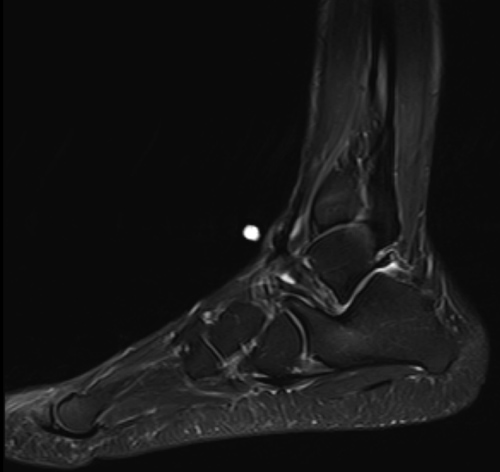
A working diagnosis of aneurysm of the dorsalis pedis artery was made as the mass was pulsatile along the dorsum of the foot.

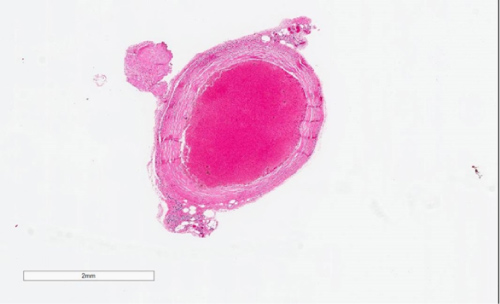
The patient was advised to opt fr an excisional biopsy so a definitive diagnosis can be made. After consent, the patient was taken to the operating room where an excisional biopsy of the soft tissue mass was performed. The sample was sent for histopathological evaluation. The pre-op course was uneventful, and so was the surgery and the post-operative period. The patient had no subsequent complications and had recovered fully till his 3-month follow-up.

Histopathological analysis revealed a diagnosis of vascularized fibroma with a blood clot inside.

Biopsy showing nodule of blood clot surrounded by fibrous wall. The lesion appears well circumscribed
SOURCEAtrushi K (2020) An Atypical Soft Tissue Mass on the Dorsum of the Foot: A Case Report. Int J Foot Ankle 4:040. doi.org/10.23937/2643-3885/1710040
Previous articleZinc Deficiency Associated Dermatitis In 4-Month-Old Boy
Next articleThe Normal Body Temperature is Dropping
Dr. Arsia Hanif has been a meritorious Healthcare professional with a proven track record throughout her academic life securing first position in her MCAT examination and then, in 2017, she successfully completed her Bachelors of Medicine and Surgery from Dow University of Health Sciences. She has had the opportunity to apply her theoretical knowledge to the real-life scenarios, as a House Officer (HO) serving at Civil Hospital. Whilst working at the Civil Hospital, she discovered that nothing satisfies her more than helping other humans in need and since then has made a commitment to implement her expertise in the field of medicine to cure the sick and regain the state of health and well-being. Being a Doctor is exactly what you’d think it’s like. She is the colleague at work that everyone wants to know but nobody wants to be. If you want to get something done, you approach her – everyone knows that! She is currently studying with Medical Council of Canada and aspires to be a leading Neurologist someday. Alongside, she has taken up medical writing to exercise her skills of delivering comprehensible version of the otherwise difficult medical literature. Her breaks comprise either of swimming, volunteering services at a Medical Camp or spending time with family.

LEAVE A REPLY

Please enter your comment!
Please enter your name here