
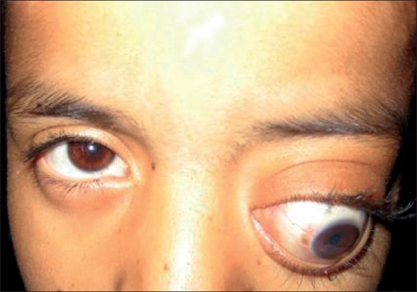
The emergency department received a 31-year-old woman with chief complaints of blurred vision in the left eye for the past 4 weeks with progressive proptosis of the left eye for the past 2 weeks.
On examination, nontender proptosis of the left eye was noticed with the inability to look laterally due to the paresis of the left abducens nerve and decreased visual acuity in the left eye.
A T2-weighted gadolinium-enhanced magnetic resonance image of the brain revealed a distinct, sharp, ovoid, cystic, and retrobulbar lesion in the left orbital cavity (Panel A). Nasal displacement of the optic nerve was noticed along with compression of the lateral rectus muscle.
Left lateral orbitotomy was performed, the cyst was separated carefully and removed. However, during the removal, the cyst ruptured. Normal saline was used to wash the area.
Histopathological evaluation revealed multiple protoscolices (Panel B, arrowheads), with central hooklets (Panel C, arrow), adjacent or attached to a thick, acellular laminated echinococcal cyst membrane (Panel B, asterisk).
The cyst was diagnosed to be an orbital hydatid cyst caused by the Echinococcus granulosus tapeworm.
Computed tomographic (CT) scans of thorax and abdomen showed no extra orbital organ involvement.
Treatment with albendazole was initiated and continued for 3 months. At 3-month follow-up, the visual acuity fully recovered.
Echinococcus granulosus tapeworm or hydatid worm infests humans when its eggs are accidentally ingested. Cysts are then formed, most commonly in the liver, but lungs and other organs may also be involved. Orbit is an extremely rare location for it, comprising only 1% of all the cystic involvements. Usually, the involvement is unilateral with no predisposition to either left or right orbit.
The most commonly observed initial clinical feature is progressive unilateral proptosis, especially if the cyst is intraorbital. Other clinical features may include loss of vision, palpebral edema, pain, redness, etc., however, in the endemic areas, if a patient presents with painless, slowly progressive proptosis, orbital hydatid cyst should top the list of differential diagnosis.
The definitive and preferred modality of treatment is the surgical removal of the cyst. However, a preoperative diagnosis with radiological investigations is strongly recommended to avoid the intraoperative rupture of the cyst.
References
Stelios F. Assimakopoulos, M. P. (2020, April 02). Orbital Hydatid Cyst. Retrieved from The New England Journal of Medicine: https://www.nejm.org/doi/full/10.1056/NEJMicm1911903
Ergün R, Okten AI, Yüksel M, et al. Orbital hydatid cysts: report of four cases. Neurosurg Rev. 1997;20:33–7.


