- This case is an unusual presentation of a ruptured Baker’s cyst in a male patient with history of ileocolonic Crohn’s disease.
- The pathology occurs as a result of fluid accumulating between the bursa of the gastrocnemius muscle and semimembranosus tendon.
- The condition is associated with other inflammatory states, for example, rheumatoid arthritis.
This case describes a 32-year-old male patient with an unusual presentation of Baker’s cyst rupture. The patient’s medical history was remarkable of ileocolonic Crohn’s disease (CD) after ileocectomy with perianal involvement, obesity, recurrent cellulitis, essential hypertension and Type 1 arthropathy with bilateral knees synovitis. The patient presented to the institution for ustekinumab therapy.
He presented with a 3-day history of right knee pain and swelling. Similarly, he reported that the pain was similar to his past arthropathy flares. He also had spondyloarthropathy, an extraintestinal manifestation of his active CD. The patient experienced intermittent fevers and chills. In addition, the pain in his knee acutely worsened during his utekinumab infusion. The pain extended into the right calf, ankle joint and foot. The patient also complained of tightness and tenderness to palpation of the posterior right calf. However, his prior Type 1 arthropathy did not present with any symptoms.
The patient’s pain had become worse upon presentation to the Emergency Department with associated nausea.
Examination and diagnosis
The affected extremity was tender to palpation with visible erythema. He did not report of any other symptoms including dizziness, lightheadedness, abdominal pain and shortness of breath. Physical examination was remarkable of tachycardia. Initial treatment included analgesics. Further tests included blood and knee joint aspiration culture. However, both were negative. Empiric antibiotic therapy was the first treatment of choice. Doctors were concerned about compartment syndrome. Therefore, they recommended consulting orthopaedics. The patient had not developed compartment syndrome. And Doppler showed his distal pulse as maintained.
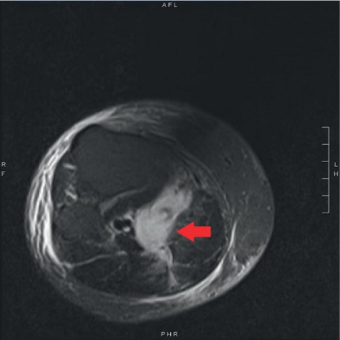
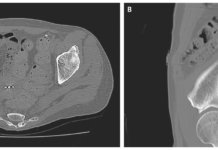
The extremity affected remained warm and well perfused. There were no significant findings on the radiographs of the lower extremity. He was evaluated for deep vein thrombosis (DVT) using ultrasound venous duplex. In addition, there were no signs of erythema and systemic symptoms. Ultrasound revealed soft tissue swelling of the posterior calf with an incidental Baker’s cyst in the calf’s medial aspect. There were also signs of small joint effusion. MRI revealed evidence of complex fluid in the right semimembranosus-gastrocnemius bursa which suggested extravasation of fluid from the Baker’s cyst rupturing. However, there were no signs of osteomyelitis. A CT of the pelvis ruled out any other causes of oedema.
Treatment
Acetaminophen and oxycodone controlled the pain well. The antibiotics were discontinued. كيف تربح في الروليت Orthopaedics recommended weight bearing and range of motion as much as the patient could tolerate. The patient was given intra-articular cortisone injections for persistent pain and swelling. Doctors continued prednisone and oral methotrexate.
The patient’s symptoms related to Baker’s cyst rupture had completely resolved at 2-month follow-up. بلاك جاك اون لاين However, he did report of persistent pain with stiffness related to his Type 1 arthropathy in the bilateral knees. Although, without any swelling. رهانات كرة القدم
References
An Unusual Presentation of Baker’s Cyst Rupture in an Inflammatory Bowel Disease Patient https://www.ncbi.nlm.nih.gov/pmc/articles/PMC6970509/


