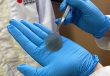

Scientists have introduced a new kind of soft 3D brain probe that may improve the way researchers record and study neural activity. Developed by researchers at Dartmouth College and partner institutions, the device is designed to better align with the brain’s natural three-dimensional structure. Traditional brain probes are usually rigid and flat, which limits their ability to capture activity occurring across different layers and regions of the brain.
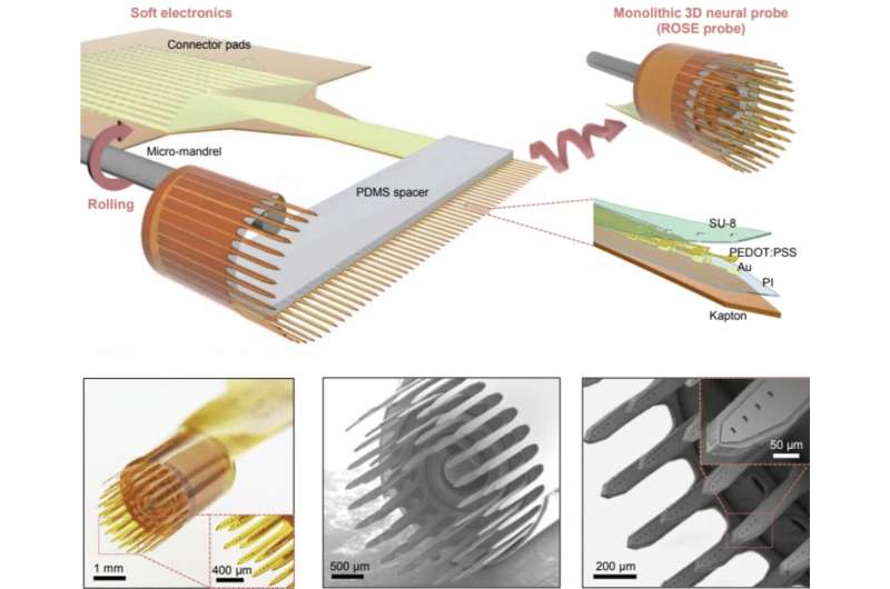
The newly developed technology, known as a ROSE probe—short for “rolling of soft electronics”—uses flexible materials that can roll into compact cylindrical forms. This design allows the probe to contain hundreds of electrodes arranged in a layered 3D configuration. By collecting signals from multiple depths at once, the device can provide a more complete picture of how neurons communicate with one another.
Researchers explained that current recording devices often fail to fully represent the complexity of brain networks because they are mainly built in two-dimensional formats. The ROSE probe addresses this limitation by creating a structure that more closely resembles the organization of brain tissue. The design has been compared to a “Swiss roll,” with multiple flexible protrusions extending into the brain while remaining compact and minimally invasive.
Another important advantage of the soft probe is its flexibility. Unlike stiff silicon-based implants, the softer material reduces irritation and inflammation in surrounding brain tissue. This may improve comfort and allow the device to function more effectively over longer periods of time.
Tests performed in awake mice and rats showed that the 3D probes were able to decode neural activity more accurately than conventional flat probes. Researchers believe the technology could eventually support advances in brain-machine interfaces, prosthetic control systems, and neuroprosthetic devices such as bionic eyes. Future studies will focus on improving long-term durability and compatibility inside living tissue.